Explore
 Validate
 Learn
Learn Western blot
 Immunocytochemistry
Antibody data
- Antibody Data
- Antigen structure
- References [4]
- Comments [0]
- Validations
- Western blot [7]
- Immunocytochemistry [1]
- Immunoprecipitation [1]
- Immunohistochemistry [3]
Submit
Validation data
Reference
Comment
Report error
- Product number
- GTX100130 - Provider product page

- Provider
- GeneTex
- Proper citation
- GeneTex Cat#GTX100130, RRID:AB_1950102
- Product name
- DDB1 antibody
- Antibody type
- Polyclonal
- Reactivity
- Human, Mouse, Rat
- Host
- Rabbit
Submitted references Human DNA Ligase I Interacts with and Is Targeted for Degradation by the DCAF7 Specificity Factor of the Cul4-DDB1 Ubiquitin Ligase Complex.
The Cullin 4A/B-DDB1-Cereblon E3 Ubiquitin Ligase Complex Mediates the Degradation of CLC-1 Chloride Channels.
Akt Phosphorylates Wnt Coactivator and Chromatin Effector Pygo2 at Serine 48 to Antagonize Its Ubiquitin/Proteasome-mediated Degradation.
DDB2 association with PCNA is required for its degradation after UV-induced DNA damage.
Peng Z, Liao Z, Matsumoto Y, Yang A, Tomkinson AE
The Journal of biological chemistry 2016 Oct 14;291(42):21893-21902
The Journal of biological chemistry 2016 Oct 14;291(42):21893-21902
The Cullin 4A/B-DDB1-Cereblon E3 Ubiquitin Ligase Complex Mediates the Degradation of CLC-1 Chloride Channels.
Chen YA, Peng YJ, Hu MC, Huang JJ, Chien YC, Wu JT, Chen TY, Tang CY
Scientific reports 2015 May 29;5:10667
Scientific reports 2015 May 29;5:10667
Akt Phosphorylates Wnt Coactivator and Chromatin Effector Pygo2 at Serine 48 to Antagonize Its Ubiquitin/Proteasome-mediated Degradation.
Li Q, Li Y, Gu B, Fang L, Zhou P, Bao S, Huang L, Dai X
The Journal of biological chemistry 2015 Aug 28;290(35):21553-67
The Journal of biological chemistry 2015 Aug 28;290(35):21553-67
DDB2 association with PCNA is required for its degradation after UV-induced DNA damage.
Cazzalini O, Perucca P, Mocchi R, Sommatis S, Prosperi E, Stivala LA
Cell cycle (Georgetown, Tex.) 2014;13(2):240-8
Cell cycle (Georgetown, Tex.) 2014;13(2):240-8
No comments: Submit comment
Enhanced validation
Supportive validation
- Submitted by
- GeneTex (provider)
- Enhanced method
- Genetic validation
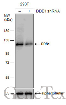
- Main image
- Experimental details
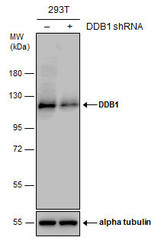
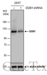
- Non-transfected (¡V) and transfected (+) 293T whole cell extracts (30 ?g) were separated by 7.5% SDS-PAGE, and the membrane was blotted with DDB1 antibody (GTX100130) diluted at 1:10000. The HRP-conjugated anti-rabbit IgG antibody (GTX213110-01) was used to detect the primary antibody.
Supportive validation
- Submitted by
- GeneTex (provider)
- Main image
- Experimental details
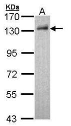
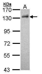
- Sample (30 ?g of whole cell lysate) A: HepG2 (GTX27900) 7.5% SDS PAGE GTX100130 diluted at 1:1000 The HRP-conjugated anti-rabbit IgG antibody (GTX213110-01) was used to detect the primary antibody.
- Submitted by
- GeneTex (provider)
- Main image

- Experimental details
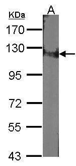
- Sample (30 ?g of whole cell lysate) A:NIH-3T37.5% SDS PAGE GTX100130 diluted at 1:1000 The HRP-conjugated anti-rabbit IgG antibody (GTX213110-01) was used to detect the primary antibody.
- Submitted by
- GeneTex (provider)
- Main image
- Experimental details
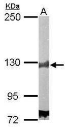
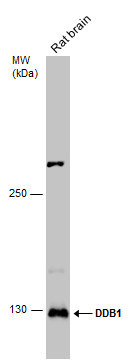
- Sample (50 ?g of whole cell lysate) A: Mouse brain 5% SDS PAGE GTX100130 diluted at 1:5000 The HRP-conjugated anti-rabbit IgG antibody (GTX213110-01) was used to detect the primary antibody.

- Submitted by
- GeneTex (provider)
- Main image
- Experimental details
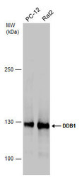
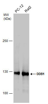
- Various whole cell extracts (30 ?g) were separated by 5% SDS-PAGE, and the membrane was blotted with DDB1 antibody (GTX100130) diluted at 1:1000. The HRP-conjugated anti-rabbit IgG antibody (GTX213110-01) was used to detect the primary antibody.
- Submitted by
- GeneTex (provider)
- Main image
- Experimental details
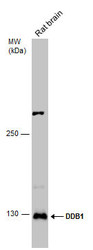
- Rat tissue extract (50 ?g) was separated by 5% SDS-PAGE, and the membrane was blotted with DDB1 antibody (GTX100130) diluted at 1:1000. The HRP-conjugated anti-rabbit IgG antibody (GTX213110-01) was used to detect the primary antibody.
- Submitted by
- GeneTex (provider)
- Main image
- Experimental details
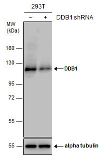
- Non-transfected (¡V) and transfected (+) 293T whole cell extracts (30 ?g) were separated by 7.5% SDS-PAGE, and the membrane was blotted with DDB1 antibody (GTX100130) diluted at 1:10000. The HRP-conjugated anti-rabbit IgG antibody (GTX213110-01) was used to detect the primary antibody.
Supportive validation
- Submitted by
- GeneTex (provider)
- Main image
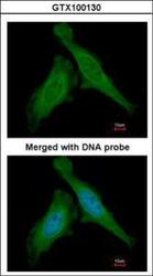
- Experimental details
- Immunofluorescence analysis of paraformaldehyde-fixed HeLa, using DDB1(GTX100130) antibody at 1:200 dilution.

Supportive validation
- Submitted by
- GeneTex (provider)
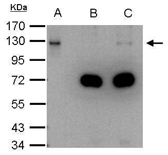
- Main image
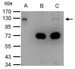
- Experimental details
- DDB1 antibody immunoprecipitates DDB1 protein in IP experiments. IP Sample: 1000 ?g HeLa whole cell lysate/extract A. 40 £gg HeLa whole cell lysate/extract B. Control with 2.5 £gg of preimmune rabbit IgG C. Immunoprecipitation of DDB1 protein by 2.5 £gg of DDB1 antibody (GTX100130) 7.5% SDS-PAGE The immunoprecipitated DDB1 protein was detected by DDB1 antibody (GTX100130) diluted at 1:1000. EasyBlot anti-rabbit IgG (GTX221666-01) was used as a secondary reagent.
Supportive validation
- Submitted by
- GeneTex (provider)
- Main image
- Experimental details
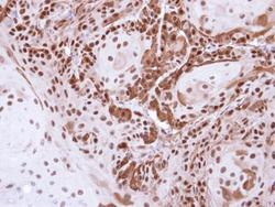
- Immunohistochemical analysis of paraffin-embedded Cal27 Xenograft , using DDB1(GTX100130) antibody at 1:500 dilution.

- Submitted by
- GeneTex (provider)
- Main image
- Experimental details
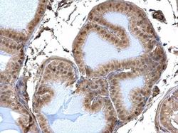
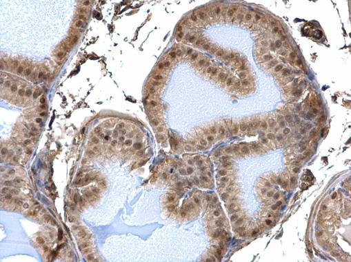
- DDB1 antibody detects DDB1 protein at nucleus and cytosol on mouse prostate by immunohistochemical analysis. Sample: Paraffin-embedded mouse prostate. DDB1 antibody (GTX100130) dilution: 1:500.
- Submitted by
- GeneTex (provider)
- Main image
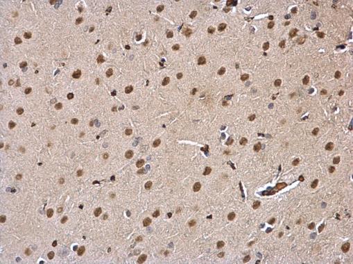
- Experimental details
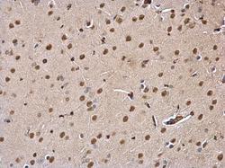
- DDB1 antibody detects DDB1 protein at nucleus and cytosol on rat fore brain by immunohistochemical analysis. Sample: Paraffin-embedded rat fore brain. DDB1 antibody (GTX100130) dilution: 1:500.