Explore
 Validate
 Learn
Learn Western blot
 ELISA
ELISA Flow cytometry
Antibody data
- Antibody Data
- Antigen structure
- References [2]
- Comments [0]
- Validations
- Flow cytometry [1]
- Other assay [4]
Submit
Validation data
Reference
Comment
Report error
- Product number
- 39-9901 - Provider product page

- Provider
- Invitrogen Antibodies
- Product name
- DDB1 Monoclonal Antibody (ZD001)
- Antibody type
- Monoclonal
- Antigen
- Recombinant full-length protein
- Reactivity
- Human, Mouse, Rat, Rabbit
- Host
- Mouse
- Isotype
- IgG
- Antibody clone number
- ZD001
- Vial size
- 100 μg
- Concentration
- 1 mg/mL
- Storage
- -20°C
Submitted references Nucleotide excision repair deficiency in melanoma in response to UVA.
Efficient parvovirus replication requires CRL4Cdt2-targeted depletion of p21 to prevent its inhibitory interaction with PCNA.
Murray HC, Maltby VE, Smith DW, Bowden NA
Experimental hematology & oncology 2015;5:6
Experimental hematology & oncology 2015;5:6
Efficient parvovirus replication requires CRL4Cdt2-targeted depletion of p21 to prevent its inhibitory interaction with PCNA.
Adeyemi RO, Fuller MS, Pintel DJ
PLoS pathogens 2014 Apr;10(4):e1004055
PLoS pathogens 2014 Apr;10(4):e1004055
No comments: Submit comment
Supportive validation
- Submitted by
- Invitrogen Antibodies (provider)
- Main image
- Experimental details
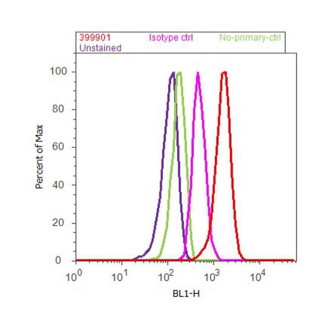
- Flow cytometry analysis of DDB1 was done on MCF7 cells. Cells were fixed with 70% ethanol for 10 minutes, permeabilized with 0.25% Triton™ X-100 for 20 minutes, and blocked with 5% BSA for 30 minutes at room temperature. Cells were labeled with DDB1 Mouse Monoclonal Antibody (Product # 39-9901, red histogram) or with mouse isotype control (pink histogram) at 3-5 µg/million cells in 2.5% BSA. After incubation at room temperature for 2 hours, the cells were labeled with Alexa Fluor® 488 Rabbit Anti-Mouse Secondary Antibody (Product # A11059) at a dilution of 1:400 for 30 minutes at room temperature. The representative 10, 000 cells were acquired and analyzed for each sample using an Attune® Acoustic Focusing Cytometer. The purple histogram represents unstained control cells and the green histogram represents no-primary-antibody control.
Supportive validation
- Submitted by
- Invitrogen Antibodies (provider)
- Main image
- Experimental details
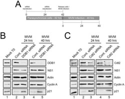
- Figure 1 p21 degradation is mediated by the CRL4 Cdt2 ligase complex. A ) Schematic illustrating the experimental protocol for siRNA knockdown of ligase components in Figures 1B and 1C . B and C ) p21 degradation requires DDB1 (B) and Cdt2 (C) . Murine A9 cells were targeted with control siRNA or siRNA to DDB1 (B) or Cdt2 (C) as depicted in Figure 1A . Uninfected control cells were harvested at the time of release (Mock T0). Infections were done at the time of release at an MOI of 10 before harvest at 24 and 40 hr pi. Western blots were performed using antibodies against the indicated proteins.

- Submitted by
- Invitrogen Antibodies (provider)
- Main image

- Experimental details
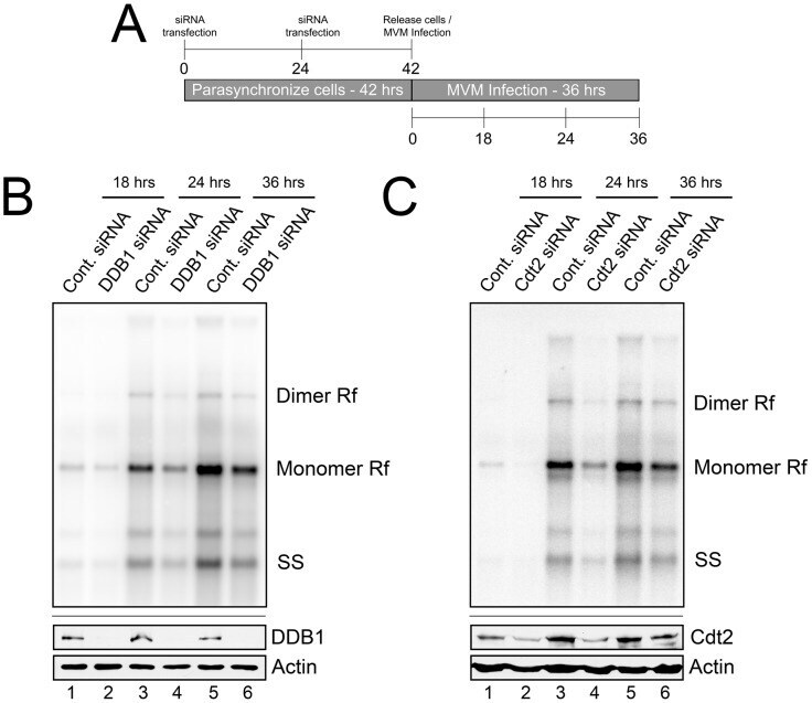
- Figure 2 The CRL4 Cdt2 E3 ligase complex is important for MVM replication. A ) Schematic showing the experimental protocol for figures 2B and 2C . B and C ) Upper panels: murine A9 cells treated with siRNA as shown in 2A were infected at an MOI of 0.5, harvested at the indicated time points and processed for Southern blotting using an MVM genomic probe. Rf - replicative forms. SS - single stranded genomic DNA. Representative Southern Blots are shown; quantifications in the text reflect two DDB1 and three Cdt2 separate knockdown experiments. Lower panels: western blots show knockdown of DDB1 and Cdt2 done in parallel experiments under identical conditions to replication assays.
- Submitted by
- Invitrogen Antibodies (provider)
- Main image
- Experimental details
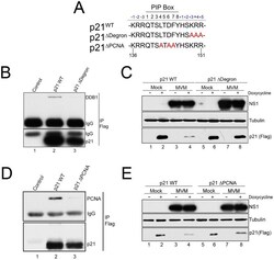
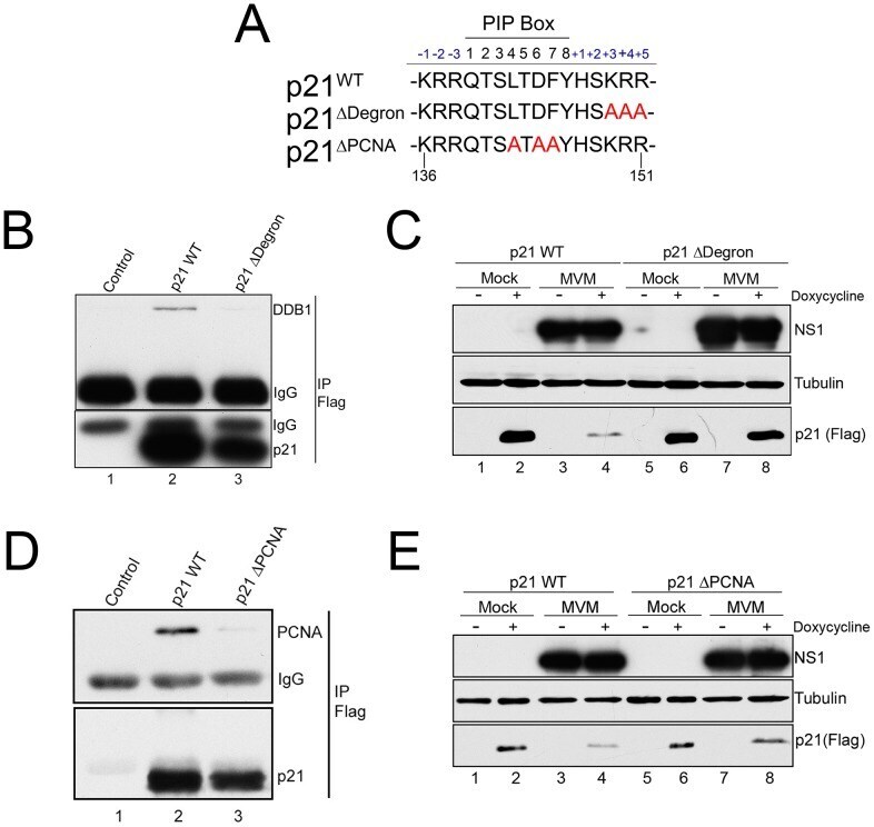
- Figure 4 p21 degradation during MVM requires interaction with PCNA and the CRL4 Cdt2 ligase complex. A ) Illustration of the p21 PIP/degron region (amino acid 136 to 151) of wild-type murine p21 (p21 WT ), p21 Deltadegron , and p21 DeltaPIP . Mutations are shown in red. B ) p21 Deltadegron does not interact with the CRL4 Cdt2 complex . 293T were cells transfected with constructs expressing FLAG-tagged p21 WT (lane 2), p21 Deltadegron (lane 3) or control plasmid (lane 1). Cells were harvested at 48 hr. Lysates were immunoprecipitated using with FLAG antibody and blotted against the indicated proteins. C ) MVM degradation of p21 requires its interaction with the CRL4 Cdt2 ligase complex . Murine A9 cell lines stably expressing p21 WT and p21 Deltadegron were generated as described. Cells were parasynchronized, released into complete media and mock-infected or infected with MVM at an MOI of 10. At 20 hr pi cells were treated with doxycycline to induce p21 expression. Cells were harvested 6 hrs later and processed for western blotting using the indicated antibodies. D ) p21 DeltaPCNA does not interact with PCNA . Experiment performed as for Figure 4B . E ) MVM degradation of p21 requires its interaction with PCNA . Experiment performed as for 4C.
- Submitted by
- Invitrogen Antibodies (provider)
- Main image
- Experimental details
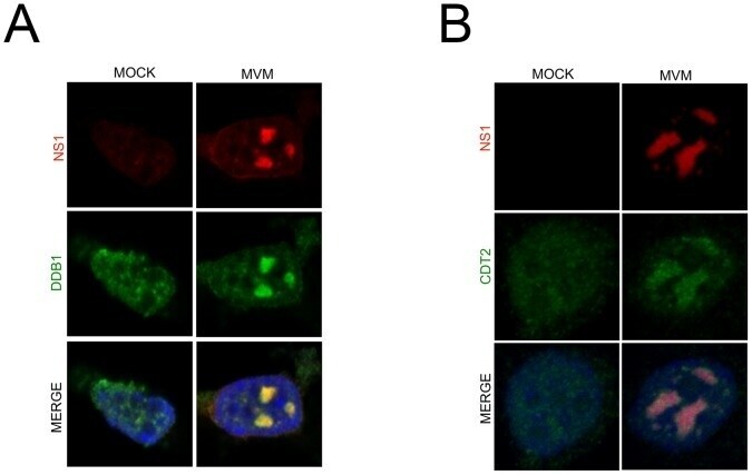
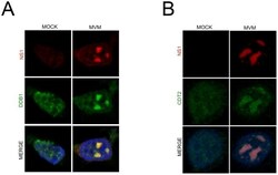
- Figure 3 The CRL4 Cdt2 ligase is recruited to viral replication compartments. A and B ) Murine A9 cells were mock infected or infected with MVM at an MOI of 10. At 24 hr pi cells were pre-extracted using cytoskeletal buffer and processed for IF using antibodies against NS1 and DDB1 (A) or Cdt2 (B).