 Explore
 Validate
 Learn
Learn Western blot
Antibody data
- Antibody Data
- Antigen structure
- References [1]
- Comments [0]
- Validations
- Western blot [3]
- Immunocytochemistry [1]
- Immunohistochemistry [4]
Submit
Validation data
Reference
Comment
Report error
- Product number
- PA5-34631 - Provider product page

- Provider
- Invitrogen Antibodies
- Product name
- DDB1 Polyclonal Antibody
- Antibody type
- Polyclonal
- Antigen
- Synthetic peptide
- Description
- Recommended positive controls: 293T, A549, H1299, HCT116, MCF-7, Neuro2A, GL261, C8D30, PC-12.
- Concentration
- 1 mg/mL
Submitted references A novel DDB2 mutation causes defective recognition of UV-induced DNA damages and prevalent equine squamous cell carcinoma.
Chen L, Bellone RR, Wang Y, Singer-Berk M, Sugasawa K, Ford JM, Artandi SE
DNA repair 2021 Jan;97:103022
DNA repair 2021 Jan;97:103022
No comments: Submit comment
Supportive validation
- Submitted by
- Invitrogen Antibodies (provider)
- Main image

- Experimental details
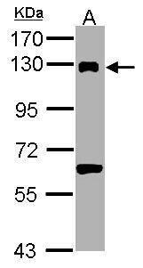
- Western Blot analysis of DDB1 was performed by separating 30 µg of PC-12 lysates by 7.5% SDS PAGE. Proteins were transferred to a membrane and probed with a DDB1 Polyclonal Antibody (Product # PA5-34631) at a dilution of 1:1000. The HRP-conjugated anti-rabbit IgG antibody was used to detect the primary antibody.
- Submitted by
- Invitrogen Antibodies (provider)
- Main image

- Experimental details
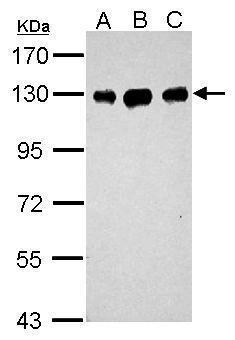
- Western Blot analysis of DDB1 was performed by separating 30 µg of various whole cell extracts by 7.5% SDS PAGE. Proteins were transferred to a membrane and probed with a DDB1 Polyclonal Antibody (Product # PA5-34631) at a dilution of 1:1000. The HRP-conjugated anti-rabbit IgG antibody was used to detect the primary antibody. A: Neuro2A, B: GL261.
- Submitted by
- Invitrogen Antibodies (provider)
- Main image

- Experimental details
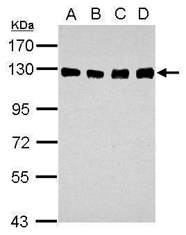
- Western Blot analysis of DDB1 was performed by separating 30 µg of various whole cell extracts by 7.5% SDSPAGE. Proteins were transferred to a membrane and probed with a DDB1 Polyclonal Antibody (Product # PA5-34631) at a dilution of 1:1000. The HRP-conjugated anti-rabbit IgG antibody was used to detect the primary antibody. A: A549, B: H1299, C: HCT116, D: MCF-7.
Supportive validation
- Submitted by
- Invitrogen Antibodies (provider)
- Main image

- Experimental details
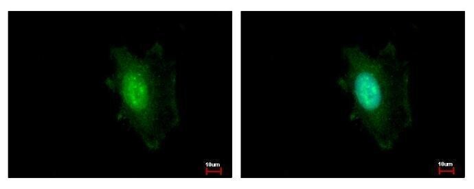
- Immunocytochemistry-Immunofluorescence analysis of DDB1 was performed in HeLa cells fixed in ice-cold MeOH for 5 min. Green: DDB1 Polyclonal Antibody (Product # PA5-34631) diluted at 1:500. Blue: Hoechst 33343 staining.
Supportive validation
- Submitted by
- Invitrogen Antibodies (provider)
- Main image
- Experimental details
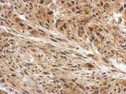
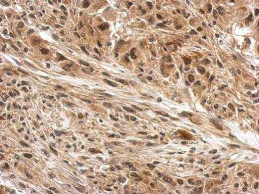
- Immunohistochemistry (Paraffin) analysis of DDB1 was performed in paraffin-embedded U373 xenograft tissue using DDB1 Polyclonal Antibody (Product # PA5-34631) at a dilution of 1:500. Antigen Retrieval: EDTA based buffer, pH 8.0, 15 min.
- Submitted by
- Invitrogen Antibodies (provider)
- Main image

- Experimental details
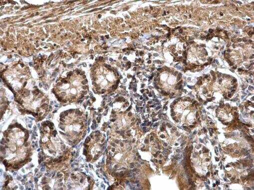
- Immunohistochemistry (Paraffin) analysis of DDB1 was performed in paraffin-embedded mouse duodenum tissue using DDB1 Polyclonal Antibody (Product # PA5-34631) at a dilution of 1:500.
- Submitted by
- Invitrogen Antibodies (provider)
- Main image

- Experimental details
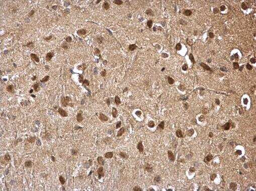
- Immunohistochemistry (Paraffin) analysis of DDB1 was performed in paraffin-embedded mouse hind brain tissue using DDB1 Polyclonal Antibody (Product # PA5-34631) at a dilution of 1:500.
- Submitted by
- Invitrogen Antibodies (provider)
- Main image
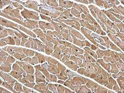
- Experimental details
- Immunohistochemistry (Paraffin) analysis of DDB1 was performed in paraffin-embedded mouse heart tissue using DDB1 Polyclonal Antibody (Product # PA5-34631) at a dilution of 1:500.
