Explore
 Validate
 Learn
Learn Western blot
Antibody data
- Antibody Data
- Antigen structure
- References [0]
- Comments [0]
- Validations
- Western blot [4]
- Immunocytochemistry [7]
- Immunohistochemistry [8]
- Flow cytometry [1]
Submit
Validation data
Reference
Comment
Report error
- Product number
- R32243 - Provider product page

- Provider
- NSJ Bioreagents
- Product name
- DDB1 Antibody
- Antibody type
- Polyclonal
- Description
- This highly specific DDB1 antibody is suitable for use in Western blot/Immunohistochemistry/Immunohistochemistry/Immunocytochemistry/Immunofluorescence/Flow cytometry applications with human, mouse and rat samples.
- Reactivity
- Human, Mouse, Rat
- Host
- Rabbit
- Conjugate
- Unconjugated
- Vial size
- 100 ug
- Concentration
- 0.5mg/ml if reconstituted with 0.2ml sterile DI water
- Storage
- After reconstitution, the DDB1 antibody can be stored for up to one month at 4oC. For long-term, aliquot and store at -20oC. Avoid repeated freezing and thawing.
No comments: Submit comment
Supportive validation
- Submitted by
- NSJ Bioreagents (provider)
- Main image

- Experimental details
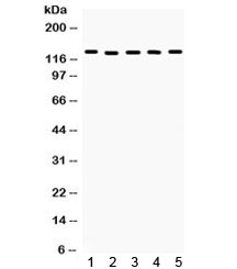
- Western blot testing of 1) rat brain, 2) rat liver, 3) mouse ovary, 4) mouse testis and 5) human MCF7 lysate. Predicted/observed molecular weight ~127 kDa.
- Submitted by
- NSJ Bioreagents (provider)
- Main image

- Experimental details
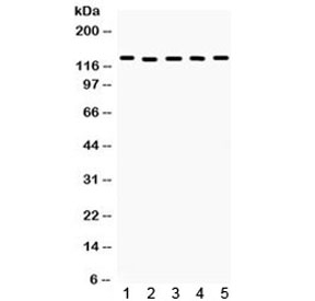
- Western blot testing of 1) rat brain, 2) rat liver, 3) mouse ovary, 4) mouse testis and 5) human MCF7 lysate. Predicted/observed molecular weight ~127 kDa.
- Submitted by
- NSJ Bioreagents (provider)
- Main image
- Experimental details
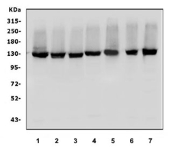
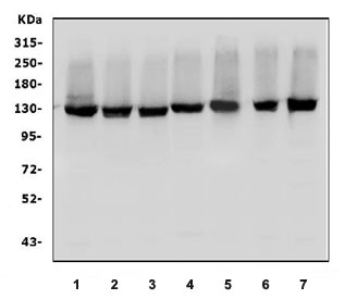
- Western blot testing of human 1) HeLa, 2) HepG2, 3) HEK293, 4) K562 5) Raji, 6) Caco-2 and 7) ThP-1 cell lysate with DDB1 antibody. Predicted molecular weight ~127 kDa.
- Submitted by
- NSJ Bioreagents (provider)
- Main image
- Experimental details
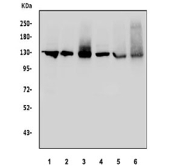
- Western blot testing of 1) rat brain, 2) rat kidney, 3) rat C6, 4) mouse brain, 5) mouse kidney and 6) mouse testis tissue lysate with DDB1 antibody. Predicted molecular weight ~127 kDa.

Supportive validation
- Submitted by
- NSJ Bioreagents (provider)
- Main image

- Experimental details
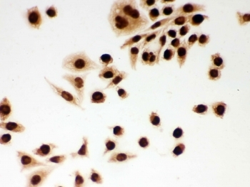
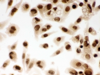
- ICC testing of FFPE human SMMC-7721 cells with DDB1 antibody. HIER: Boil the paraffin sections in pH 6, 10mM citrate buffer for 20 minutes and allow to cool prior to staining.
- Submitted by
- NSJ Bioreagents (provider)
- Main image
- Experimental details
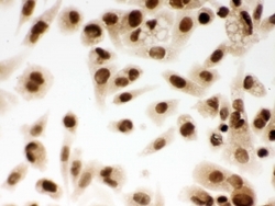
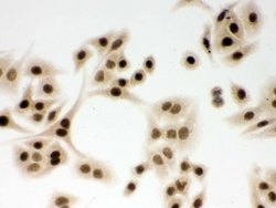
- ICC testing of FFPE human PC-3 cells with DDB1 antibody. HIER: Boil the paraffin sections in pH 6, 10mM citrate buffer for 20 minutes and allow to cool prior to staining.
- Submitted by
- NSJ Bioreagents (provider)
- Main image
- Experimental details
- ICC testing of FFPE human A549 cells with DDB1 antibody. HIER: Boil the paraffin sections in pH 6, 10mM citrate buffer for 20 minutes and allow to cool prior to staining.

- Submitted by
- NSJ Bioreagents (provider)
- Main image
- Experimental details
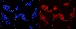
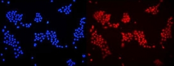
- Immunofluorescent staining of FFPE human A431 cells with DDB1 antibody (red) and DAPI nuclear stain (blue). HIER: steam section in pH6 citrate buffer for 20 min.
- Submitted by
- NSJ Bioreagents (provider)
- Main image
- Experimental details
- ICC testing of FFPE human SMMC-7721 cells with DDB1 antibody. HIER: Boil the paraffin sections in pH 6, 10mM citrate buffer for 20 minutes and allow to cool prior to staining.

- Submitted by
- NSJ Bioreagents (provider)
- Main image

- Experimental details
- ICC testing of FFPE human PC-3 cells with DDB1 antibody. HIER: Boil the paraffin sections in pH 6, 10mM citrate buffer for 20 minutes and allow to cool prior to staining.

- Submitted by
- NSJ Bioreagents (provider)
- Main image

- Experimental details
- ICC testing of FFPE human A549 cells with DDB1 antibody. HIER: Boil the paraffin sections in pH 6, 10mM citrate buffer for 20 minutes and allow to cool prior to staining.

Supportive validation
- Submitted by
- NSJ Bioreagents (provider)
- Main image

- Experimental details
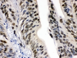
- IHC testing of FFPE human intestinal cancer with DDB1 antibody. HIER: Boil the paraffin sections in pH 6, 10mM citrate buffer for 20 minutes and allow to cool prior to staining.

- Submitted by
- NSJ Bioreagents (provider)
- Main image

- Experimental details
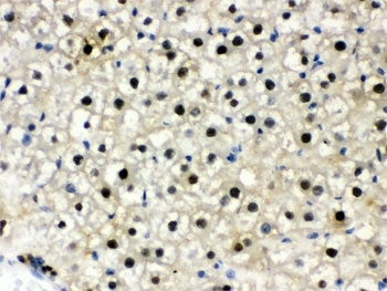
- IHC testing of FFPE mouse liver with DDB1 antibody. HIER: Boil the paraffin sections in pH 6, 10mM citrate buffer for 20 minutes and allow to cool prior to staining.

- Submitted by
- NSJ Bioreagents (provider)
- Main image
- Experimental details
- IHC testing of FFPE rat liver with DDB1 antibody. HIER: Boil the paraffin sections in pH 6, 10mM citrate buffer for 20 minutes and allow to cool prior to staining.
- Submitted by
- NSJ Bioreagents (provider)
- Main image
- Experimental details
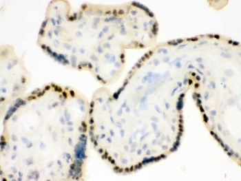
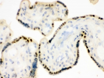
- IHC testing of frozen human placental tissue with DDB1 antibody.
- Submitted by
- NSJ Bioreagents (provider)
- Main image
- Experimental details
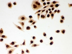
- IHC testing of FFPE human intestinal cancer with DDB1 antibody. HIER: Boil the paraffin sections in pH 6, 10mM citrate buffer for 20 minutes and allow to cool prior to staining.

- Submitted by
- NSJ Bioreagents (provider)
- Main image

- Experimental details
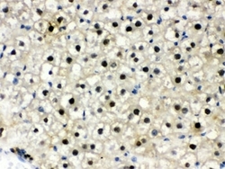
- IHC testing of FFPE mouse liver with DDB1 antibody. HIER: Boil the paraffin sections in pH 6, 10mM citrate buffer for 20 minutes and allow to cool prior to staining.

- Submitted by
- NSJ Bioreagents (provider)
- Main image

- Experimental details
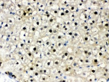
- IHC testing of FFPE rat liver with DDB1 antibody. HIER: Boil the paraffin sections in pH 6, 10mM citrate buffer for 20 minutes and allow to cool prior to staining.
- Submitted by
- NSJ Bioreagents (provider)
- Main image

- Experimental details
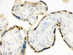
- IHC testing of frozen human placental tissue with DDB1 antibody.
Supportive validation
- Submitted by
- NSJ Bioreagents (provider)
- Main image
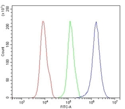
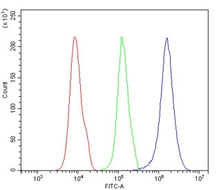
- Experimental details
- Flow cytometry testing of human 293T cells with DDB1 antibody at 1ug/million cells (blocked with goat sera); Red=cells alone, Green=isotype control, Blue= DDB1 antibody.