Explore
 Validate
 Learn
Learn Western blot
 ELISA
ELISAAntibody data
- Antibody Data
- Antigen structure
- References [1]
- Comments [0]
- Validations
- Western blot [1]
- Immunocytochemistry [2]
Submit
Validation data
Reference
Comment
Report error
- Product number
- GTX48825 - Provider product page

- Provider
- GeneTex
- Proper citation
- GeneTex Cat#GTX48825, RRID:AB_11178438
- Product name
- Keratin antibody [C11]
- Antibody type
- Monoclonal
- Reactivity
- Human
- Host
- Mouse
Submitted references Broad-spectrum immunohistochemical epithelial markers: a review.
Ordóñez NG
Human pathology 2013 Jul;44(7):1195-215
Human pathology 2013 Jul;44(7):1195-215
No comments: Submit comment
Supportive validation
- Submitted by
- GeneTex (provider)
- Main image
- Experimental details
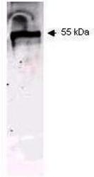
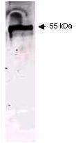
- Western blot using GeneTex Immunochemical's Mouse Mab-anti-Keratin antibody. This antibody recognizes a single 56 kDa band corresponding to human keratin as confirmed by the position of molecular weight markers (not shown). Approximatley 100 µg of keratin from human epidermis (Sigma p/n K0253) was applied under reducing conditions to a pre-cast 4-20% µgel from Gradipore Inc. A 1:400 dilution of Mab anti-Keratin was used for 2h followed by detection using a 1:5,000 dilution of IRDyeTM800 conjugated Goat anti-Mouse IgG [H&L] and visualization using the Odyssey® Infrared Imaging System developed by LI-COR. Other detection systems will yield similar results. IRDye is a trademark of LI-COR, Inc.
- Validation comment
- WB
Supportive validation
- Submitted by
- GeneTex (provider)
- Main image
- Experimental details
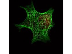
- Immunofluorescence Microscopy of GeneTex Anti-Keratin antibody (GTX48825) was used with GeneTex Dylight 488 goat anti-mouse (shown in green) to detect Keratin by Immunofluorescence. In the same experiment, GeneTex polyclonal Anti-HDAC-1 antibody was used with Anti-Rabbit IgG (shown in red) to detect HDAC-1.

- Submitted by
- GeneTex (provider)
- Main image

- Experimental details
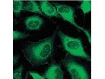
- Immunofluorescence microscopy using GeneTex Immunochemical's Mouse Mab-anti-Keratin antibody. Confocal slices of HeLa cells are between 0.5 and 0.6 um where the image is taken near the bottom of the cell. Use FITC a 1:2,000 dilution of FITC-conjμgated Goat anti-Mouse IgG [H&L] for detection.