Explore
 Validate
 Learn
LearnMA5-14965
antibody from Invitrogen Antibodies
Targeting: TCF7
TCF-1
 Western blot Immunocytochemistry
 Immunoprecipitation Immunohistochemistry Flow cytometry Other assay
Antibody data
- Antibody Data
- Antigen structure
- References [4]
- Comments [0]
- Validations
- Western blot [3]
- Immunocytochemistry [2]
- Immunohistochemistry [1]
- Other assay [3]
Submit
Validation data
Reference
Comment
Report error
- Product number
- MA5-14965 - Provider product page

- Provider
- Invitrogen Antibodies
- Product name
- TCF7 Monoclonal Antibody (C.725.7)
- Antibody type
- Monoclonal
- Antigen
- Synthetic peptide
- Description
- It is not recommended to aliquot this antibody. This antibody is not cross-reactive with LEF1.
- Reactivity
- Human, Mouse
- Host
- Rabbit
- Isotype
- IgG
- Antibody clone number
- C.725.7
- Vial size
- 100 μL
- Concentration
- 100 μg/mL
- Storage
- -20°C
Submitted references Oncogenic cooperation between TCF7-SPI1 and NRAS(G12D) requires β-catenin activity to drive T-cell acute lymphoblastic leukemia.
Chronic expression of p16(INK4a) in the epidermis induces Wnt-mediated hyperplasia and promotes tumor initiation.
Intrinsic 4-1BB signals are indispensable for the establishment of an influenza-specific tissue-resident memory CD8 T-cell population in the lung.
β-catenin is required for taste bud cell renewal and behavioral taste perception in adult mice.
Van Thillo Q, De Bie J, Seneviratne JA, Demeyer S, Omari S, Balachandran A, Zhai V, Tam WL, Sweron B, Geerdens E, Gielen O, Provost S, Segers H, Boeckx N, Marshall GM, Cheung BB, Isobe K, Kato I, Takita J, Amos TG, Deveson IW, McCalmont H, Lock RB, Oxley EP, Garwood MM, Dickins RA, Uyttebroeck A, Carter DR, Cools J, de Bock CE
Nature communications 2021 Jul 6;12(1):4164
Nature communications 2021 Jul 6;12(1):4164
Chronic expression of p16(INK4a) in the epidermis induces Wnt-mediated hyperplasia and promotes tumor initiation.
Azazmeh N, Assouline B, Winter E, Ruppo S, Nevo Y, Maly A, Meir K, Witkiewicz AK, Cohen J, Rizou SV, Pikarsky E, Luxenburg C, Gorgoulis VG, Ben-Porath I
Nature communications 2020 Jun 1;11(1):2711
Nature communications 2020 Jun 1;11(1):2711
Intrinsic 4-1BB signals are indispensable for the establishment of an influenza-specific tissue-resident memory CD8 T-cell population in the lung.
Zhou AC, Wagar LE, Wortzman ME, Watts TH
Mucosal immunology 2017 Sep;10(5):1294-1309
Mucosal immunology 2017 Sep;10(5):1294-1309
β-catenin is required for taste bud cell renewal and behavioral taste perception in adult mice.
Gaillard D, Bowles SG, Salcedo E, Xu M, Millar SE, Barlow LA
PLoS genetics 2017 Aug;13(8):e1006990
PLoS genetics 2017 Aug;13(8):e1006990
No comments: Submit comment
Supportive validation
- Submitted by
- Invitrogen Antibodies (provider)
- Main image

- Experimental details
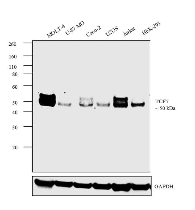
- Western blot analysis was performed on whole cell extracts (30 µg lysate) of MOLT-4 (Lane 1), U-87 MG (Lane 2), Caco-2 (Lane 3), U2OS (Lane 4), Jurkat (Lane 5) and HEK-293 (Lane 6). The blot was probed with anti-TCF7 Monoclonal Antibody (Product # MA5-14965,1:1000 dilution) and detected by chemiluminescence using Goat anti-Rabbit IgG (Heavy Chain) Superclonal™ Secondary Antibody, HRP conjugate (Product # A27036, 0.25 µg/mL, 1:4000 dilution). A 50 kDa band corresponding to TCF7 was observed across the cell lines tested.
- Submitted by
- Invitrogen Antibodies (provider)
- Main image
- Experimental details
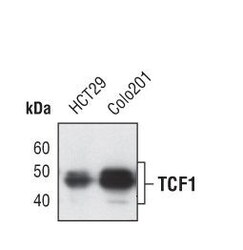
- Western blot analysis of TCF1 in total cell lysates from HCT29 and Colo201 cells using TCF1 monoclonal antibody (Product # MA5-14965).

- Submitted by
- Invitrogen Antibodies (provider)
- Main image
- Experimental details
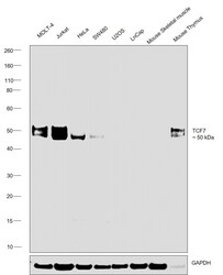
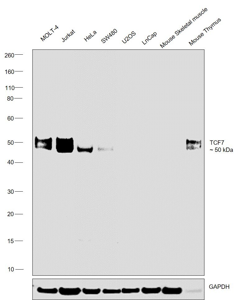
- Western blot was performed using Anti-TCF7 Monoclonal Antibody (C.725.7) (Product # MA5-14965) and a 50 kDa band corresponding to TCF7 was observed across across the cell lines and tissues tested. Whole cell extracts (30 µg lysate) of MOLT-4 (Lane 1), Jurkat (Lane 2), HeLa (Lane 3), SW480 (Lane 4), U-2 OS (Lane 5), LNCaP (Lane 6), Mouse Skeletal Muscle (Lane 7), Mouse Thymus (Lane 8) were electrophoresed using NuPAGE™ 4-12% Bis-Tris Protein Gel (Product # NP0322BOX). Resolved proteins were then transferred onto a nitrocellulose membrane (Product # IB23001) by iBlot® 2 Dry Blotting System (Product # IB21001). The blot was probed with the primary antibody (1:1000 dilution) and detected by chemiluminescence with Goat anti-Mouse IgG (H+L) Superclonal™ Recombinant Secondary Antibody, HRP (Product # A28177,1:20000 dilution) using the iBright™ FL1500 Imaging System (Product # A44115). Chemiluminescent detection was performed using SuperSignal™ West Atto Ultimate Sensitivity Substrate (Product # A38556).
Supportive validation
- Submitted by
- Invitrogen Antibodies (provider)
- Main image

- Experimental details
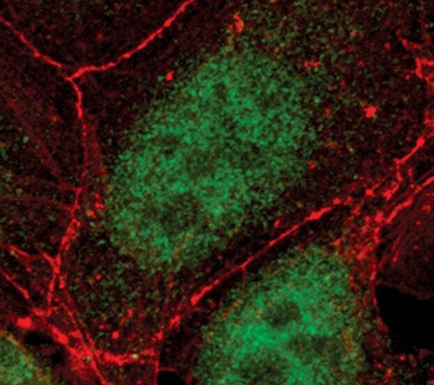
- Immunofluorescent analysis of TCF1 in DLD-1 cells using a TCF1 monoclonal antibody (Product # MA5-14965) (green). Actin filaments are labeled with a fluorescent red phalloidin.
- Submitted by
- Invitrogen Antibodies (provider)
- Main image

- Experimental details
- Immunofluorescent analysis of TCF1 in DLD-1 cells using a TCF1 monoclonal antibody (Product # MA5-14965) (green). Actin filaments are labeled with a fluorescent red phalloidin.

Supportive validation
- Submitted by
- Invitrogen Antibodies (provider)
- Main image
- Experimental details
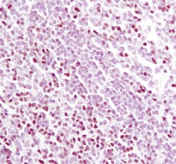
- Immunohistochemical analysis of TCF1 in paraffin-embedded human tonsil using a TCF1 monoclonal antibody (Product # MA5-14965).

Supportive validation
- Submitted by
- Invitrogen Antibodies (provider)
- Main image
- Experimental details
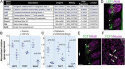
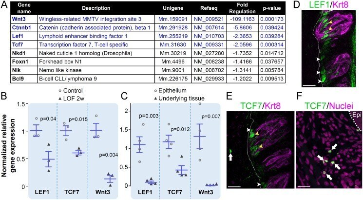
- 10.1371/journal.pgen.1006990.g006 Fig 6 LEF1, TCF7 and Wnt3 are downregulated in Krt5-beta-catenin LOF CVP. To identify Wnt pathway genes regulated in the absence of beta-catenin, a Wnt pathway RT 2 profiler PCR assay was run on RNAs extracted from whole CVP of Krt5-beta-catenin LOF mice and their control counterparts (see Methods ). As expected, beta-catenin expression was downregulated 6 fold in Krt5-beta-catenin LOF mice compared with controls ( A ). Seven other genes were significantly regulated; all being downregulated ( A ) and attention was focused on genes which expression changed more than 2 fold, i . e . LEF1, TCF7 and Wnt3 ( A , genes in blue). Regulation of LEF1, TCF7 and Wnt3 expression was confirmed by qRT-PCR ( B ). qRT-PCR analysis of epithelium versus underlying tissue (mesenchyme and Von Ebner's glands) of wild-type CVPs revealed that LEF1 and Wnt3 are specifically expressed in the epithelium whereas expression of TCF7 is predominant in the epithelium and lower in the underlying tissue ( C ). Immunolabelling of LEF1 and TCF7 is localized in perigemmal basal cell, which include progenitors ( D , E , white arrowheads) and within taste buds ( D , E , yellow arrowheads). In addition, TCF7 signal was observed in the mesenchyme ( E , F , white arrows) supporting the qRT-PCR data. Dotted line delineates the basement membrane; Epi: CVP trench epithelium. N = 3 mice per group, except C (n = 4). Data are represented as scatter plots (individual symbols), and mean +- S
- Submitted by
- Invitrogen Antibodies (provider)
- Main image
- Experimental details
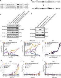
- Fig. 3 The presence of the NRAS (G12D) mutation is necessary for in vitro transformation to cytokine-independent growth. a Constitutive retroviral vectors with either BFP-P2A-TCF7SPI1, BFP-P2A-NRAS(G12D) or TCF7-SPI1-P2A-NRAS(G12D) followed by an IRES-sequence and GFP. b Schematic representation of the inducible Cre-Lox system. The same constructs as in ( a ) were cloned in the antisense direction between 2 anti-parallel asymmetric Lox66/Lox71 sites. Upon expression of Cre-recombinase the sequence is flipped in a unidirectional manner. c , d Western blot analysis showing expression of TCF7, SPI1, and NRAS for the different indicated constructs in Ba/F3 cells ( c ) ( n = 1) or Cre-Ba/F3 cells with active Cre-recombinase ( d ) ( n = 1). WT = Wild-type Ba/F3 and used as a control lysate. The viral P2A sequences within the expression vector results in a 3' in-frame 18 amino acid sequence tail on the first protein expressed accounting for the increased molecular weight for NRAS(G12D) and TCF7-SPI1 in ( c ) and ( d ), respectively. e Growth curve of Ba/F3 cells transduced with the vectors illustrated in ( a ) or empty vector (white). f Growth curve with floxed constructs illustrated in ( b ) in Ba/F3-Cre cells alongside an empty vector control. g - j Growth curves in pro-T cells after transduction with the indicated constructs and empty vector. Either no growth factors ( g ), Scf ( h ), Il7 ( i ) or Dll4 ( j ) were omitted. e - j The number of cells is shown as a mean with standard
- Submitted by
- Invitrogen Antibodies (provider)
- Main image
- Experimental details
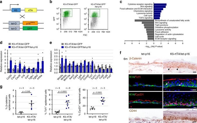
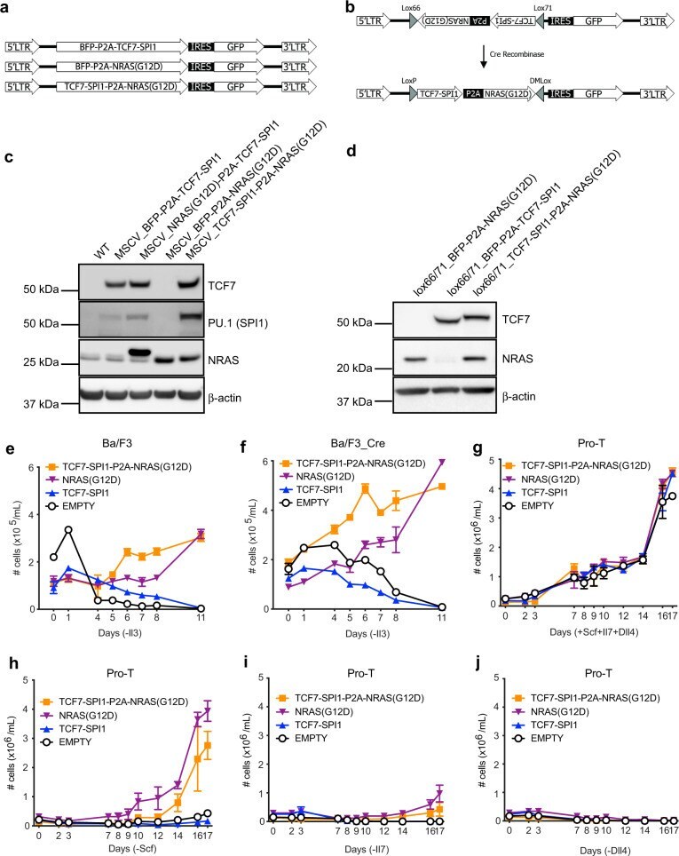
- Fig. 4 p16 expression activates Wnt-pathway-associated genes. a Diagram of transgenic mouse lines crossed for co-induction of GFP and p16. b FACS plots showing GFP levels and gates used for isolation of GFP + epidermal cells from K5-rtTA/tet-GFP/tet-p16 (right) and control K5-rtTA/tet-GFP (left) mice, after 6 months of induction. Plots show the CD31 - /CD140a - /CD45 - cell fraction only. c Gene sets whose expression was preferentially upregulated (blue) or downregulated (grey) in GFP + cells from p16-expressing mice relative to GFP + cells from control mice. Values indicate -log 10 (adjusted P value) by hypergeometric test. d mRNA levels of genes encoding cytokines and genes associated with the TGFbeta pathway upregulated in p16-expressing GFP + cells, measured by mRNA-seq. Values were normalized to the mean expression levels in GFP + cells from control mice, defined as 1. P < 0.05 for all genes, n = 4 samples per group, each pooled from 3 mice. e Relative mRNA levels of genes associated with the Wnt pathway in the same samples. P < 0.05 for all genes, n = 4 samples per group, each pooled from 3 mice. f Skin sections from K5-rtTA/tet-p16 and control tet-p16 mice treated with dox for 6 months stained for beta-Catenin, Tcf1, Cyclin D1 or CD44. Arrowheads indicate representative positively stained cells. g Percentages of nuclear beta-catenin + , Tcf1 + , and CyclinD1 + IFE cells in control and p16-expressing mice after 6 months of induction, as scored visually from images. P va