 Explore
 Validate
 Learn
Learn Western blot
 Immunocytochemistry
 Immunoprecipitation
Antibody data
- Antibody Data
- Antigen structure
- References [2]
- Comments [0]
- Validations
- Immunocytochemistry [3]
- Flow cytometry [1]
- Other assay [3]
Submit
Validation data
Reference
Comment
Report error
- Product number
- PA5-17551 - Provider product page

- Provider
- Invitrogen Antibodies
- Product name
- PKC alpha Polyclonal Antibody
- Antibody type
- Polyclonal
- Antigen
- Synthetic peptide
- Description
- Antibodies to this protein (and modification) were previously sold as part of a Thermo Scientific Cellomics High Content Screening Kit. This replacement antibody is now recommended for researchers who need an antibody for high content cell based assays. It has been thoroughly tested and validated for cellular immunofluorescence (IF) applications. Further optimization including the selection of the most appropriate fluorescent Dylight conjugated secondary antibody may have to be performed for your high content assay. It is not recommended to aliquot this antibody. This antibody is not cross-reactive with endogenous levels of other PKC isoforms.
- Reactivity
- Human, Mouse, Rat
- Host
- Rabbit
- Isotype
- IgG
- Vial size
- 100 µL
- Concentration
- 11 µg/mL
- Storage
- -20°C
Submitted references Cell signaling pathways in autosomal-dominant leukodystrophy (ADLD): the intriguing role of the astrocytes.
A New Mouse Model Related to SCA14 Carrying a Pseudosubstrate Domain Mutation in PKCγ Shows Perturbed Purkinje Cell Maturation and Ataxic Motor Behavior.
Ratti S, Rusciano I, Mongiorgi S, Owusu Obeng E, Cappellini A, Teti G, Falconi M, Talozzi L, Capellari S, Bartoletti-Stella A, Guaraldi P, Cortelli P, Suh PG, Cocco L, Manzoli L, Ramazzotti G
Cellular and molecular life sciences : CMLS 2021 Mar;78(6):2781-2795
Cellular and molecular life sciences : CMLS 2021 Mar;78(6):2781-2795
A New Mouse Model Related to SCA14 Carrying a Pseudosubstrate Domain Mutation in PKCγ Shows Perturbed Purkinje Cell Maturation and Ataxic Motor Behavior.
Shimobayashi E, Kapfhammer JP
The Journal of neuroscience : the official journal of the Society for Neuroscience 2021 Mar 3;41(9):2053-2068
The Journal of neuroscience : the official journal of the Society for Neuroscience 2021 Mar 3;41(9):2053-2068
No comments: Submit comment
Supportive validation
- Submitted by
- Invitrogen Antibodies (provider)
- Main image

- Experimental details
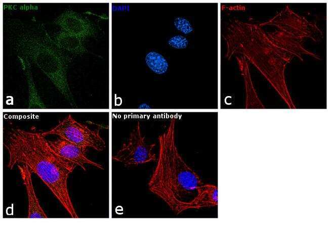
- Immunofluorescence analysis of PKC alpha was performed using 70% confluent log phase NIH/3T3 cells. The cells were fixed with 4% paraformaldehyde for 10 minutes, permeabilized with 0.1% Triton™ X-100 for 10 minutes, and blocked with 2% BSA for 1 hour at room temperature. The cells were labeled with PKC alpha Polyclonal Antibody (Product # PA5-17551) at 1:100 dilution in 0.1% BSA and incubated overnight at 4 degree and then labeled with Goat anti-Rabbit IgG (H+L) Superclonal™ Secondary Antibody, Alexa Fluor® 488 conjugate (Product # A27034) at a dilution of 1:2000 for 45 minutes at room temperature (Panel a: green). Nuclei (Panel b: blue) were stained with SlowFade® Gold Antifade Mountant with DAPI (Product # S36938). F-actin (Panel c: red) was stained with Rhodamine Phalloidin (Product # R415, 1:300). Panel d represents the merged image showing cytoplasmic localization. Panel e represents control cells with no primary antibody to assess background. The images were captured at 60X magnification.
- Submitted by
- Invitrogen Antibodies (provider)
- Main image

- Experimental details
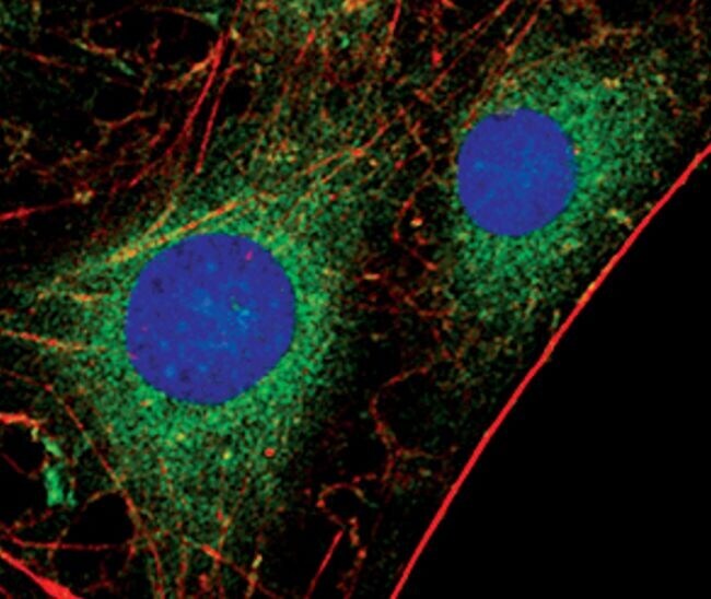
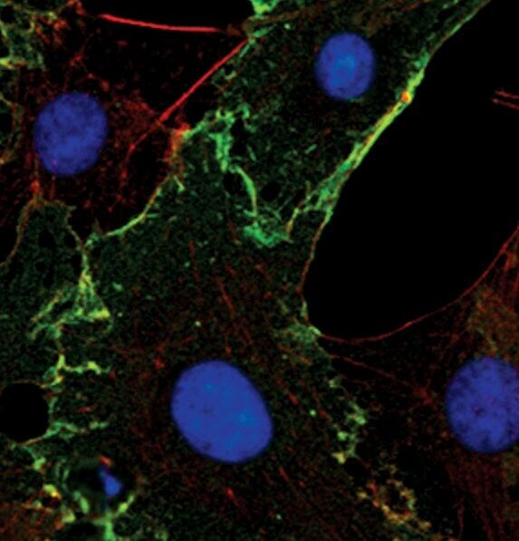
- Immunofluorescent analysis of PKC-alpha in C6 cells, TPA-treated, using a PKC-alpha polyclonal antibody (Product # PA5-17551) (green). Actin filaments are labeled with a fluorescent red phalloidin. DNA is labeled using a fluorescent blue dye.
- Submitted by
- Invitrogen Antibodies (provider)
- Main image
- Experimental details
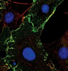
- Immunofluorescent analysis of PKC-alpha in C6 cells, serum-starved, using a PKC-alpha polyclonal antibody (Product # PA5-17551) (green). Actin filaments are labeled with a fluorescent red phalloidin. DNA is labeled using a fluorescent blue dye.
Supportive validation
- Submitted by
- Invitrogen Antibodies (provider)
- Main image
- Experimental details
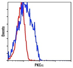
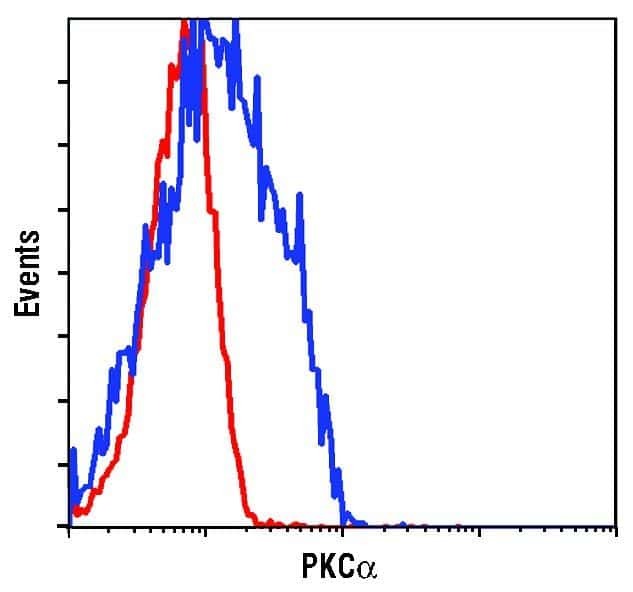
- Flow cytometric analysis of PKC-alpha in untreated 293 cells using a PKC-alpha polyclonal antibody (Product # PA5-17551) (blue) compared to a nonspecific negative control antibody (red).
Supportive validation
- Submitted by
- Invitrogen Antibodies (provider)
- Main image
- Experimental details
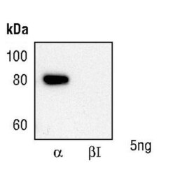
- Antibody specificity is demonstrated by a recombinant protein array. Western blotting of recombinant human PKC alpha and Beta proteins using PKC alpha Antibody (Product # PA5-17551) demonstrates isoform-specificity of the PKC-alpha antibody.

- Submitted by
- Invitrogen Antibodies (provider)
- Main image
- Experimental details
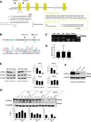
- Figure 3. Generation and characterization of the PKC gamma -A24E mouse. A , Target sequence in the PRKCG gene (chromosome 7, 1.93 cM) for producing point mutated (c. 71C > A; p. Ala 24 Glu) PRKCG with Cas9/CRISPR engineering system. To prevent donor DNA cleavage, the PAM sequence mutation was also introduced (78G > A), which does not give a change in amino acids. B , Mutations were validated with sequencing. The figure shows Het PKC gamma -A24E sequence. C , To identify the genotypes, PCR was performed followed by Alu1 (BioLabs, R0137S) digestion for 1 h, which digests only Ala 24 Glu mutated DNA. Wt shows a single band (250 bp), Het PKC gamma -A24E shows a Wt band and two digested bands (250, 150, and 100 bp), and Homo PKC gamma -A24E shows only two digested bands (150 and 100 bp). D , qPCR using 5-week-old cerebellar samples from each genotype. Data show that all three genotypes express PKCgamma mRNA. E , Western blot analysis of total PKCgamma protein in the cerebellum from the three genotypes ( n = 4). PKCgamma protein degradation in PKC gamma -A24E mice was observed (4-week-old: Wt = 100.0 +- 8.34%, n = 4; Het = 49.12 +- 6.80%, n = 4; Homo = 7.519 +- 2.48%, p = 0.0286, n = 4; 6-week-old: Wt = 100.0 +- 14.56%, n = 4; Het = 63.09 +- 6.39%, n = 4; Homo = 4.674 +- 0.40%, p = 0.0286, n = 4), whereas PKCalpha protein expression did not change in all genotypes (4-week-old: Wt = 100.0 +- 9.30%, n = 4; Het = 83.13 +- 2.09%, n = 4; Homo = 87.67 +- 14.1%, n = 4; 6-week-old: Wt = 10

- Submitted by
- Invitrogen Antibodies (provider)
- Main image
- Experimental details
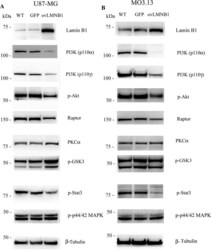
- Fig. 3 Lamin B1 build up affects the expression and phosphorylation of signaling molecules downstream LIF-R. The expression and the phosphorylation of molecules downstream LIF- signaling pathway was evaluated in U87-MG ( a ) and MO3.13 ( b ) cells 96 h after transduction. Lamin B1 overexpressing cells ( ovLMNB1 ) were compared to wild type ( WT ) and mock-transduced ( GFP ) samples. The expression or phosphorylation of the denoted proteins was assayed. beta-tubulin was used as loading control. Western blot results are representative of three independent experiments
