 Explore
 Validate
 Learn
Learn Western blot
 Immunocytochemistry
Antibody data
- Antibody Data
- Antigen structure
- References [2]
- Comments [0]
- Validations
- Immunocytochemistry [1]
Submit
Validation data
Reference
Comment
Report error
- Product number
- MAB2786 - Provider product page

- Provider
- R&D Systems
- Product name
- Mouse IL-24 Antibody
- Antibody type
- Monoclonal
- Description
- Protein A or G purified from hybridoma culture supernatant. Detects mouse IL-24 in direct ELISAs and Western blots. No cross-reactivity with recombinant human IL-24 is observed.
- Reactivity
- Mouse
- Host
- Rat
- Conjugate
- Unconjugated
- Antigen sequence
NP_444325- Isotype
- IgG
- Antibody clone number
- 303308
- Vial size
- 100 ug
- Concentration
- LYOPH
- Storage
- Use a manual defrost freezer and avoid repeated freeze-thaw cycles. 12 months from date of receipt, -20 to -70 °C as supplied. 1 month, 2 to 8 °C under sterile conditions after reconstitution. 6 months, -20 to -70 °C under sterile conditions after reconstitution.
Submitted references Murine astrocytes produce IL-24 and are susceptible to the immunosuppressive effects of this cytokine.
Dysregulation of suppressor of cytokine signaling 3 in keratinocytes causes skin inflammation mediated by interleukin-20 receptor-related cytokines.
Burmeister AR, Johnson MB, Yaemmongkol JJ, Marriott I
Journal of neuroinflammation 2019 Mar 2;16(1):55
Journal of neuroinflammation 2019 Mar 2;16(1):55
Dysregulation of suppressor of cytokine signaling 3 in keratinocytes causes skin inflammation mediated by interleukin-20 receptor-related cytokines.
Uto-Konomi A, Miyauchi K, Ozaki N, Motomura Y, Suzuki Y, Yoshimura A, Suzuki S, Cua D, Kubo M
PloS one 2012;7(7):e40343
PloS one 2012;7(7):e40343
No comments: Submit comment
Supportive validation
- Submitted by
- R&D Systems (provider)
- Main image
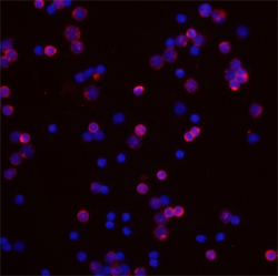
- Experimental details
- IL-24 in Mouse Splenocytes. IL-24 was detected in immersion fixed mouse splenocytes using Rat Anti-Mouse IL-24 Monoclonal Antibody (Catalog # MAB2786) at 10 µg/mL for 3 hours at room temperature. Cells were stained using the Northern-Lights™ 557-conjugated Anti-Rat IgG Secondary Antibody (red; Catalog # NL013) and counterstained with DAPI (blue). Specific staining was localized to cytoplasm. View our protocol for Fluorescent ICC Staining of Non-adherent Cells.
