Explore
 Validate
 Learn
Learn Immunohistochemistry
Antibody data
- Antibody Data
- Antigen structure
- References [0]
- Comments [0]
- Validations
- Immunohistochemistry [3]
Submit
Validation data
Reference
Comment
Report error
- Product number
- 56-9003-82 - Provider product page

- Provider
- Invitrogen Antibodies
- Product name
- Pan Cytokeratin Monoclonal Antibody (AE1/AE3), Alexa Fluor™ 700
- Antibody type
- Monoclonal
- Antigen
- Other
- Description
- Description: The monoclonal antibodies AE1 and AE3 recognize many of the acidic and basic cytokeratin family members. Cytokeratins are intermediate filament proteins comprising one component of the cytoskeleton. There are two large families of cytokeratins, acidic and basic, but all contain the same basic domains (i.e. an alpha-helical core with an N- and C-terminal domain). The proteins are expressed in epithelial cells, but are developmentally regulated. Many tumors also express these proteins and their expression can help identify the origin of a neoplasm. The AE3 monoclonal antibody recognizes the 65 to 67 triplet, 64, 59, 58, 56, 54 and 52kD proteins also known as cytokeratin 1, 2, 3, 4, 5, 6, 7, and 8 while the AE1 antibody recognizes 56.5, 54', 50, 50', 48, and 40 kDa proteins (also known as CK10, 14, 15, 16 and 19). These antibodies can be used on a wide array of tissue samples from mouse, human, rat, primates (cynomolgus and rhesus), dog, cat, rabbit, and chicken. Applications Reported: This AE1/AE3 antibody has been reported for use in immunohistochemical staining, immunocytochemistry, immunohistochemical staining of frozen tissue sections, and immunohistochemical staining of formalin-fixed paraffin embedded tissue sections. Applications Tested: This AE1/AE3 antibody has been tested by immunohistochemistry of formalin-fixed paraffin embedded tissue using high pH antigen retrieval conditions and can be used at 2 µg/mL. It is recommended that the antibody be carefully titrated for optimal performance in the assay of interest. Using conjugate solutions: Centrifuge the protein conjugate solution briefly in a microcentrifuge before use; add only the supernatant to the experiment. This step will help eliminate any protein aggregates that may have formed during storage, thereby reducing nonspecific background staining.
- Reactivity
- Human
- Host
- Mouse
- Conjugate
- Near infrared dye
- Isotype
- IgG
- Antibody clone number
- AE1/AE3
- Vial size
- 100 μg
- Concentration
- 0.2 mg/mL
- Storage
- 4°C, store in dark, DO NOT FREEZE!
No comments: Submit comment
Supportive validation
- Submitted by
- Invitrogen Antibodies (provider)
- Main image
- Experimental details
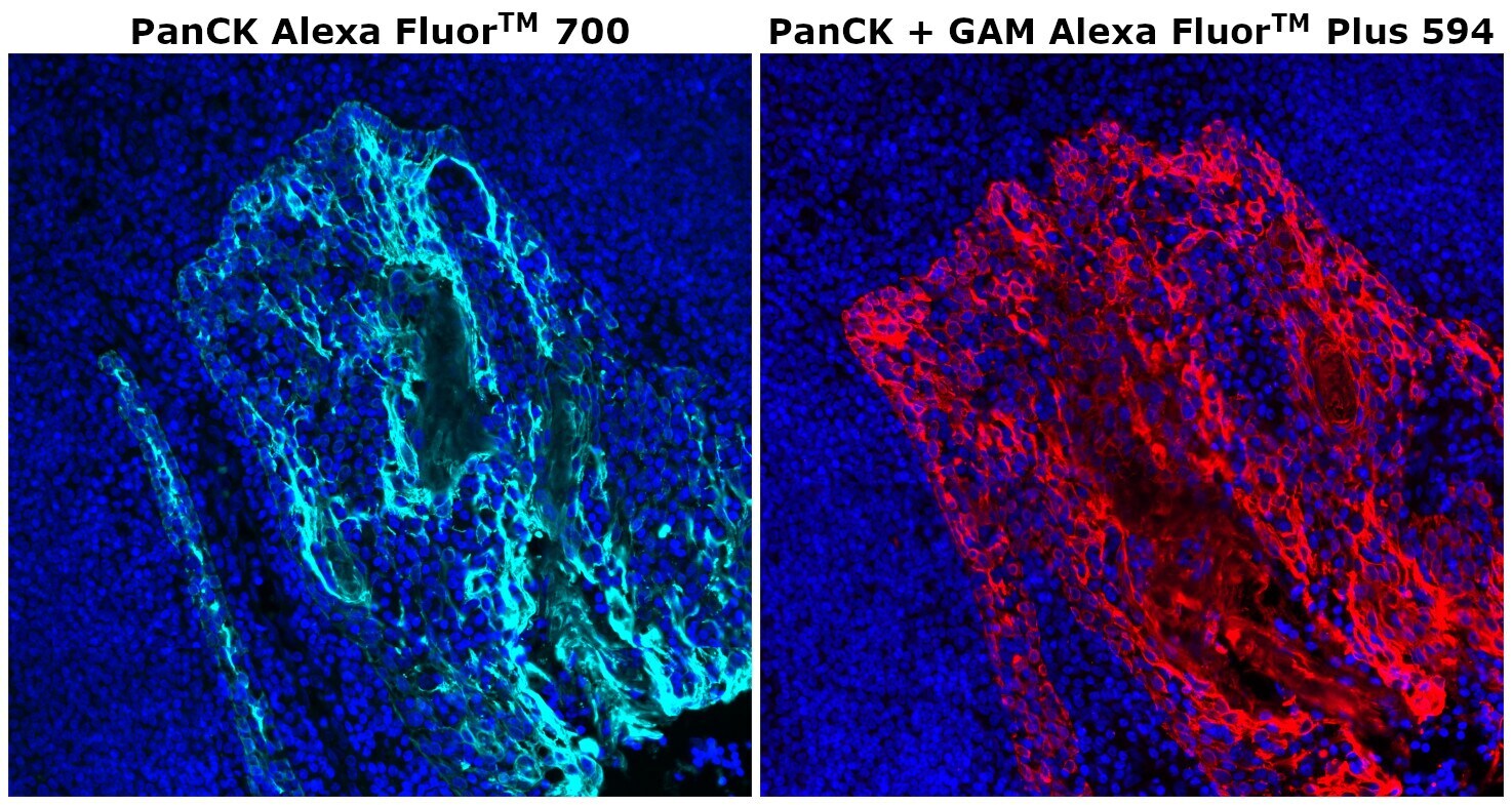
- Immunohistochemical analysis of PanCK was performed on FFPE human tonsil tissue. To expose the target protein, HIER was performed on de-paraffinized sections using BOND Epitope Retrieval Solution 2 (pH 9) for 10 mins, followed by a 5-min cool down and a 5-min wash with ddH2O. Tissues were permeabilized with 0.1% Triton X-100 in 1X PBS for 30 mins and blocked with 3% BSA/5% normal goat serum/1X PBS for 1 hr at RT. Following the removal of the blocking solution, tissues were probed with Pan Cytokeratin Monoclonal Antibody (AE1/AE3), Alexa Fluor™ 700 (Product # 56-9003-82) at 2 µg/mL (left) or Cytokeratin Pan Type I/II Antibody Cocktail (AE1/AE3), eBioscience™ (Product # MA5-13156) at 0.5 µg/mL (right), respectively, in blocking solution for one hr at RT in a humidified chamber. Tissues were then washed three times, 5 mins each, in 1X PBS. Detection of the unconjugated primary antibody was performed using Goat anti-Mouse IgG (H+L) Highly Cross-Adsorbed Secondary Antibody, Alexa Fluor™ Plus 594 (Product # A32742) at a dilution of 1:2,000 in blocking solution for one hr at RT, followed by three, 5-minute washes in 1X PBS. Sections were stained with DAPI (Product # 62247) at 1 µg/mL diluted in 1X PBS for 5 minutes at RT, washed twice in 1X PBS followed by a final rinse in ddH2O. Tissues were mounted with ProLong™ Glass Antifade Mountant (Product # P36982) and images were captured on EVOS™ M7000 Imaging System (Product # AMF7000) at 20X magnification.
- Submitted by
- Invitrogen Antibodies (provider)
- Main image
- Experimental details
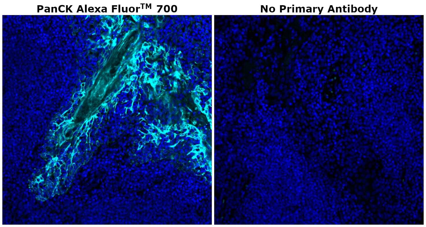
- Immunohistochemical analysis of PanCK was performed on formalin-fixed paraffin-embedded human tonsil tissue. To expose the target protein, heat-induced epitope retrieval (HIER) was performed on de-paraffinized sections using BOND Epitope Retrieval Solution 2 (pH 9) diluted to 1X solution in water in a pressure cooker for ten minutes followed by a 5-minute cool down and a 5-minute wash with ddH2O. Tissues were permeabilized with 0.1% Triton X-100 in 1X PBS for 30 minutes and blocked with 3% BSA/5% normal goat serum/1X PBS for 1 hour at room temperature. Following the removal of the blocking solution, tissues were probed with (left) or without (right) Pan Cytokeratin Monoclonal Antibody (AE1/AE3), Alexa Fluor™ 700 (Product # 56-9003-82) at 2 µg/mL in blocking solution for one hour at room temperature in a humidified chamber. Tissues were then washed three times, 5 minutes each, in 1X PBS and then stained with DAPI (Product # P36982) at 1 µg/mL diluted in 1X PBS for 5 minutes at room temperature. The sections were washed twice in 1X PBS and followed by a final rinse in ddH2O and mounted with ProLong™ Glass Antifade Mountant (Product # P36982) and images were captured on EVOS™ M7000 Imaging System (Product # AMF7000) at 20X magnification.
- Submitted by
- Invitrogen Antibodies (provider)
- Main image
- Experimental details
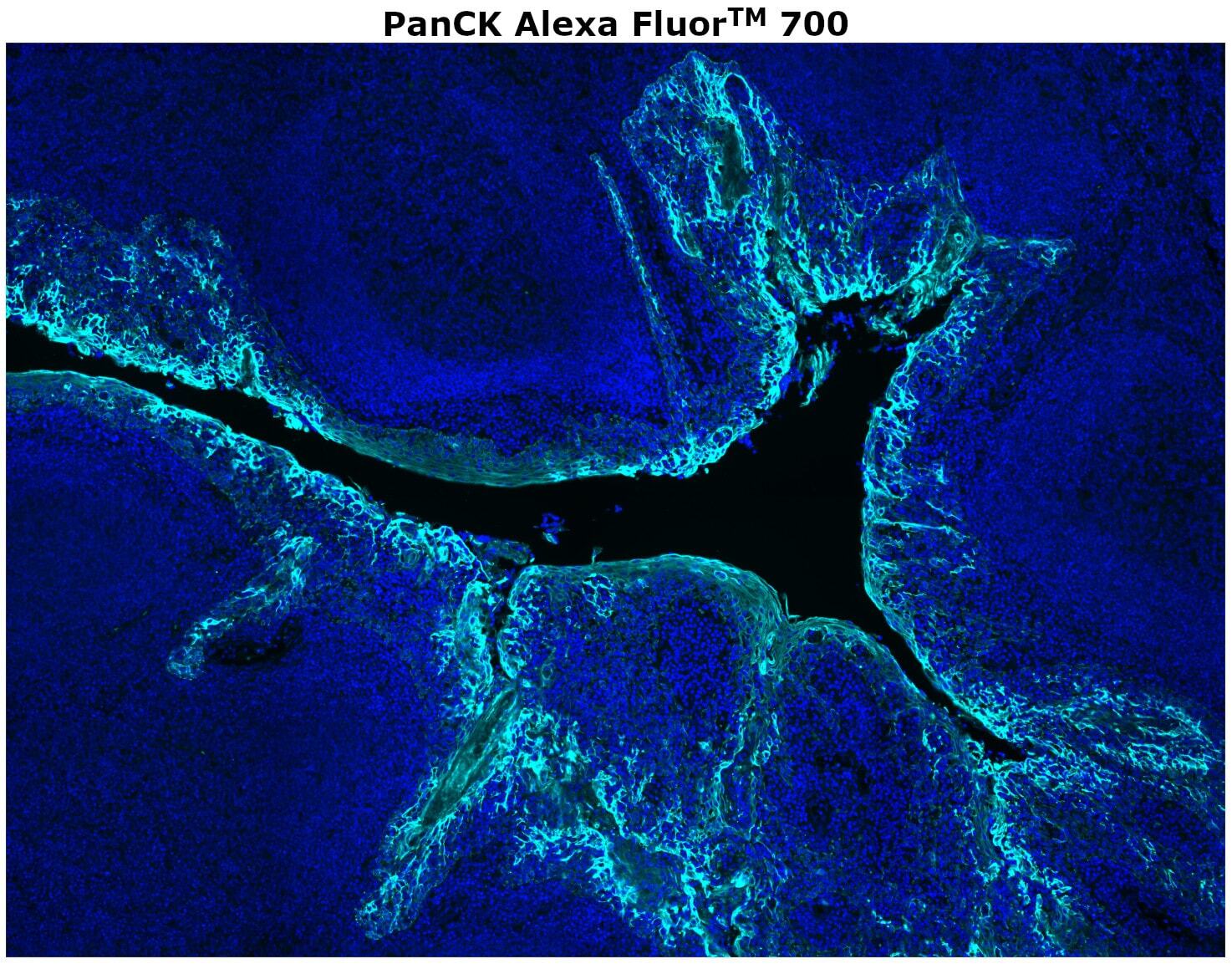
- Immunohistochemical analysis of PanCK was performed on formalin-fixed paraffin-embedded human tonsil tissue. To expose the target protein, heat-induced epitope retrieval (HIER) was performed on de-paraffinized sections using BOND Epitope Retrieval Solution 2 (pH 9) for 10 minutes, followed by a 5-minute cool down and a 5-minute wash with ddH2O. The tissue was then permeabilized with 0.1% Triton X-100 in 1X PBS for 30 minutes and blocked with 3% BSA/5% normal goat serum/1X PBS for 1 hour at room temperature. Following the removal of the blocking solution, the tissue was probed with Pan Cytokeratin Monoclonal Antibody (AE1/AE3), Alexa Fluor™ 700 (Product # 56-9003-82) at 2 µg/mL in blocking solution for one hour at room temperature in a humidified chamber. The tissue was then washed three times, 5 minutes each, in 1X PBS and then stained with DAPI (Product # P36982) at 1 µg/mL diluted in 1X PBS for 5 minutes at room temperature. The section was washed twice in 1X PBS followed by a final rinse in ddH2O and mounted with ProLong™ Glass Antifade Mountant (Product # P36982) and the image was captured on EVOS™ M7000 Imaging System (Product # AMF7000) at 20X magnification.