 Explore
 Validate
 Learn
Learn Western blot
Antibody data
- Antibody Data
- Antigen structure
- References [8]
- Comments [0]
- Validations
- Western blot [1]
- Immunohistochemistry [1]
- Flow cytometry [1]
Submit
Validation data
Reference
Comment
Report error
- Product number
- MA5-13150 - Provider product page

- Provider
- Invitrogen Antibodies
- Product name
- Cytokeratin Pan Type II Monoclonal Antibody (AE3)
- Antibody type
- Monoclonal
- Antigen
- Other
- Description
- MA5-13150 targets Cytokeratin High Molecular Weight in IF and IHC (P) applications and shows reactivity with Bovine, Chicken, Human, mouse, Non-human primate, Rabbit, and Rat samples.
- Antibody clone number
- AE3
- Concentration
- 0.2 mg/mL
Submitted references Novel immunofluorescence protocol for multimarker assessment of putative disseminating breast cancer stem cells.
Assessment of the tumorigenesis and drug susceptibility of three new canine mammary tumor cell lines.
An immunohistochemical study of feline endometrial adenocarcinoma.
Coexistence of different tissue tumourigenesis in an N-methyl-N-nitrosourea-induced mammary carcinoma model: a histopathological report in Sprague-Dawley rats.
BMP4 signaling regulates formation of Hertwig's epithelial root sheath during tooth root development.
Graft-versus-host-like disease complicating thymoma: lack of AIRE expression as a cause of non-hereditary autoimmunity?
Mediastinal intraoperative radioisotope sentinel lymph node mapping in non-small-cell lung cancer.
Cytokeratin expression in lichen amyloidosus and macular amyloidosis.
Balic M, Rapp N, Stanzer S, Lin H, Strutz J, Szkandera J, Daidone MG, Samonigg H, Cote RJ, Dandachi N
Applied immunohistochemistry & molecular morphology : AIMM 2011 Jan;19(1):33-40
Applied immunohistochemistry & molecular morphology : AIMM 2011 Jan;19(1):33-40
Assessment of the tumorigenesis and drug susceptibility of three new canine mammary tumor cell lines.
Chang CY, Chiou PP, Chen WJ, Li YH, Yiu JC, Cheng YH, Chen SD, Lin CT, Lai YS
Research in veterinary science 2010 Apr;88(2):285-93
Research in veterinary science 2010 Apr;88(2):285-93
An immunohistochemical study of feline endometrial adenocarcinoma.
Gil da Costa RM, Santos M, Amorim I, Lopes C, Pereira PD, Faustino AM
Journal of comparative pathology 2009 May;140(4):254-9
Journal of comparative pathology 2009 May;140(4):254-9
Coexistence of different tissue tumourigenesis in an N-methyl-N-nitrosourea-induced mammary carcinoma model: a histopathological report in Sprague-Dawley rats.
Esendagli G, Yilmaz G, Canpinar H, Gunel-Ozcan A, Guc MO, Guc D
Laboratory animals 2009 Jan;43(1):60-4
Laboratory animals 2009 Jan;43(1):60-4
BMP4 signaling regulates formation of Hertwig's epithelial root sheath during tooth root development.
Hosoya A, Kim JY, Cho SW, Jung HS
Cell and tissue research 2008 Sep;333(3):503-9
Cell and tissue research 2008 Sep;333(3):503-9
Graft-versus-host-like disease complicating thymoma: lack of AIRE expression as a cause of non-hereditary autoimmunity?
Offerhaus GJ, Schipper ME, Lazenby AJ, Montgomery E, Morsink FH, Bende RJ, Musler AR, van Lier RA, van Noesel CJ
Immunology letters 2007 Nov 30;114(1):31-7
Immunology letters 2007 Nov 30;114(1):31-7
Mediastinal intraoperative radioisotope sentinel lymph node mapping in non-small-cell lung cancer.
Atinkaya C, Ozlem Küçük N, Koparal H, Aras G, Sak SD, Ozdemir N
Nuclear medicine communications 2005 Aug;26(8):717-20
Nuclear medicine communications 2005 Aug;26(8):717-20
Cytokeratin expression in lichen amyloidosus and macular amyloidosis.
Apaydin R, Gürbüz Y, Bayramgürler D, Müezzinoglu B, Bilen N
Journal of the European Academy of Dermatology and Venereology : JEADV 2004 May;18(3):305-9
Journal of the European Academy of Dermatology and Venereology : JEADV 2004 May;18(3):305-9
No comments: Submit comment
Supportive validation
- Submitted by
- Invitrogen Antibodies (provider)
- Main image

- Experimental details
- Western blot analysis was performed on whole cell extracts of T- 47D (Lane 1), PC-12 (Lane 2) and MCF7 (Lane 3). The blot was probed with anti-Cytokeratin Pan Type II Mouse Monoclonal Antibody (Product # MA5-13150, 2 µg/mL) and detected by chemiluminescence using Goat anti-Mouse IgG (H+L) Superclonal™ Secondary Antibody, HRP conjugate (Product # A28177, 0.4 µg/mL, 1:2500 dilution). Bands between ~ 40-70 kDa corresponding to Cytokeratin Pan Type II were observed across cell lines tested. Known quantity of protein samples were electrophoresed using Novex® NuPAGE® 4-12 % Bis-Tris gel (Product # NP0321BOX), XCell SureLock™ Electrophoresis System (Product # EI0002) and Novex® Sharp Pre-Stained Protein Standard (Product # LC5800). Resolved proteins were then transferred onto a nitrocellulose membrane with iBlot® 2 Dry Blotting System (Product # IB21001). The membrane was probed with the relevant primary and secondary Antibody following blocking with 5 % skimmed milk. Chemiluminescent detection was performed using Pierce™ ECL Western Blotting Substrate (Product # 32106).

Supportive validation
- Submitted by
- Invitrogen Antibodies (provider)
- Main image

- Experimental details
- Formalin-fixed, paraffin-embedded human skin stained with Keratin, HMW antibody using peroxidase-conjugate and AEC chromogen. Note cytoplasmic staining of epithelial cells.

Supportive validation
- Submitted by
- Invitrogen Antibodies (provider)
- Main image
- Experimental details
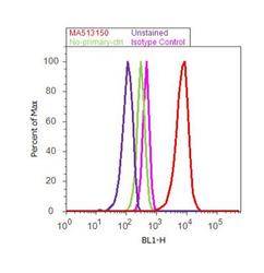
- Flow cytometry analysis of Cytokeratin Pan Type II was done on MCF7 cells. Cells were fixed with 70% ethanol for 10 minutes, permeabilized with 0.25% Triton™ X-100 for 20 minutes, and blocked with 5% BSA for 30 minutes at room temperature. Cells were labeled with Cytokeratin Pan Type II Mouse Monoclonal Antibody (MA5-13150, red histogram) or with mouse isotype control (pink histogram) at 3-5 ug/million cells in 2.5% BSA. After incubation at room temperature for 2 hours, the cells were labeled with Alexa Fluor® 488 Rabbit Anti-Mouse Secondary Antibody (A11059) at a dilution of 1:400 for 30 minutes at room temperature. The representative 10, 000 cells were acquired and analyzed for each sample using an Attune® Acoustic Focusing Cytometer. The purple histogram represents unstained control cells and the green histogram represents no-primary-antibody control.
