Explore
 Validate
 Learn
Learn Western blot
 Flow cytometry
Antibody data
- Antibody Data
- Antigen structure
- References [6]
- Comments [0]
- Validations
- Flow cytometry [1]
Submit
Validation data
Reference
Comment
Report error
- Product number
- PA1-734 - Provider product page

- Provider
- Invitrogen Antibodies
- Product name
- SCAMP2 Polyclonal Antibody
- Antibody type
- Polyclonal
- Antigen
- Synthetic peptide
- Description
- PA1-734 detects secretory carrier membrane protein 2 (SCAMP 2) from mouse testes and AtT20 cell extract as well as Chinese hamster ovary (CHO) cell extract. PA1-734 has been successfully used in Western blot procedures. By Western blot, this antibody detects an ~38 kDa protein representing SCAMP 2 from CHO cell extract. PA1-734 immunizing peptide corresponds to amino acid residues 1-17 from human SCAMP 2. This sequence is completely conserved between human and mouse. PA1-734 immunizing peptide (Cat. # PEP-151) is available for use in neutralization and control experiments.
- Reactivity
- Human, Mouse, Hamster
- Host
- Rabbit
- Isotype
- IgG
- Vial size
- 100 μg
- Concentration
- 1 mg/mL
- Storage
- -20°C, Avoid Freeze/Thaw Cycles
Submitted references Tissue expression of the vesicle protein pantophysin.
Tissue expression of the vesicle protein pantophysin.
Tyrosine phosphorylation of selected secretory carrier membrane proteins, SCAMP1 and SCAMP3, and association with the EGF receptor.
Tyrosine phosphorylation of selected secretory carrier membrane proteins, SCAMP1 and SCAMP3, and association with the EGF receptor.
Three mammalian SCAMPs (secretory carrier membrane proteins) are highly related products of distinct genes having similar subcellular distributions.
Three mammalian SCAMPs (secretory carrier membrane proteins) are highly related products of distinct genes having similar subcellular distributions.
Windoffer R, Borchert-Stuhlträger M, Haass NK, Thomas S, Hergt M, Bulitta CJ, Leube RE
Cell and tissue research 1999 Jun;296(3):499-510
Cell and tissue research 1999 Jun;296(3):499-510
Tissue expression of the vesicle protein pantophysin.
Windoffer R, Borchert-Stuhlträger M, Haass NK, Thomas S, Hergt M, Bulitta CJ, Leube RE
Cell and tissue research 1999 Jun;296(3):499-510
Cell and tissue research 1999 Jun;296(3):499-510
Tyrosine phosphorylation of selected secretory carrier membrane proteins, SCAMP1 and SCAMP3, and association with the EGF receptor.
Wu TT, Castle JD
Molecular biology of the cell 1998 Jul;9(7):1661-74
Molecular biology of the cell 1998 Jul;9(7):1661-74
Tyrosine phosphorylation of selected secretory carrier membrane proteins, SCAMP1 and SCAMP3, and association with the EGF receptor.
Wu TT, Castle JD
Molecular biology of the cell 1998 Jul;9(7):1661-74
Molecular biology of the cell 1998 Jul;9(7):1661-74
Three mammalian SCAMPs (secretory carrier membrane proteins) are highly related products of distinct genes having similar subcellular distributions.
Singleton DR, Wu TT, Castle JD
Journal of cell science 1997 Sep;110 ( Pt 17):2099-107
Journal of cell science 1997 Sep;110 ( Pt 17):2099-107
Three mammalian SCAMPs (secretory carrier membrane proteins) are highly related products of distinct genes having similar subcellular distributions.
Singleton DR, Wu TT, Castle JD
Journal of cell science 1997 Sep;110 ( Pt 17):2099-107
Journal of cell science 1997 Sep;110 ( Pt 17):2099-107
No comments: Submit comment
Supportive validation
- Submitted by
- Invitrogen Antibodies (provider)
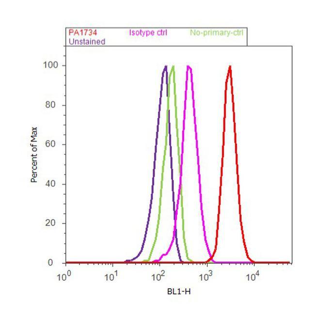
- Main image
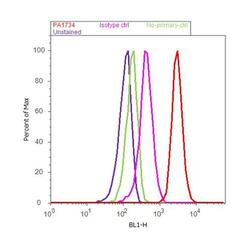
- Experimental details
- Flow cytometry analysis of SCAMP2 was done on F9 cells. Cells were fixed with 70% ethanol for 10 minutes, permeabilized with 0.25% Triton™ X-100 for 20 minutes, and blocked with 5% BSA for 30 minutes at room temperature. Cells were labeled with SCAMP2 Rabbit Polyclonal Antibody (PA1734, red histogram) or with rabbit isotype control (pink histogram) at 3-5 ug/million cells in 2.5% BSA. After incubation at room temperature for 2 hours, the cells were labeled with Alexa Fluor® 488 Goat Anti-Rabbit Secondary Antibody (A11008) at a dilution of 1:400 for 30 minutes at room temperature. The representative 10,000 cells were acquired and analyzed for each sample using an Attune® Acoustic Focusing Cytometer. The purple histogram represents unstained control cells and the green histogram represents no-primary-antibody control..