 Explore
 Validate
 Learn
Learn Western blot
 Immunohistochemistry
Antibody data
- Antibody Data
- Antigen structure
- References [4]
- Comments [0]
- Validations
- Western blot [1]
- Immunocytochemistry [2]
Submit
Validation data
Reference
Comment
Report error
- Product number
- HPA016737 - Provider product page

- Provider
- Atlas Antibodies
- Proper citation
- Atlas Antibodies Cat#HPA016737, RRID:AB_1857271
- Product name
- Anti-SMARCAD1
- Antibody type
- Polyclonal
- Description
- Polyclonal Antibody against Human SMARCAD1, Gene description: SWI/SNF-related, matrix-associated actin-dependent regulator of chromatin, subfamily a, containing DEAD/H box 1, Alternative Gene Names: DKFZP762K2015, ETL1, KIAA1122, Validated applications: ICC, IHC, WB, Uniprot ID: Q9H4L7, Storage: Store at +4°C for short term storage. Long time storage is recommended at -20°C.
- Reactivity
- Human, Mouse, Rat
- Host
- Rabbit
- Conjugate
- Unconjugated
- Isotype
- IgG
- Vial size
- 100 µl
- Concentration
- 0.6 mg/ml
- Storage
- Store at +4°C for short term storage. Long time storage is recommended at -20°C.
- Handling
- The antibody solution should be gently mixed before use.
Submitted references The Conserved Chromatin Remodeler SMARCAD1 Interacts with TFIIIC and Architectural Proteins in Human and Mouse
Smarcad1 mediates microbiota-induced inflammation in mouse and coordinates gene expression in the intestinal epithelium
An embryonic stem cell-specific heterochromatin state promotes core histone exchange in the absence of DNA accessibility
The CUE1 domain of the SNF2-like chromatin remodeler SMARCAD1 mediates its association with KRAB-associated protein 1 (KAP1) and KAP1 target genes
Sachs P, Bergmaier P, Treutwein K, Mermoud J
Genes 2023;14(9):1793
Genes 2023;14(9):1793
Smarcad1 mediates microbiota-induced inflammation in mouse and coordinates gene expression in the intestinal epithelium
Kazakevych J, Denizot J, Liebert A, Portovedo M, Mosavie M, Jain P, Stellato C, Fraser C, Corrêa R, Célestine M, Mattiuz R, Okkenhaug H, Miller J, Vinolo M, Veldhoen M, Varga-Weisz P
Genome Biology 2020;21(1)
Genome Biology 2020;21(1)
An embryonic stem cell-specific heterochromatin state promotes core histone exchange in the absence of DNA accessibility
Navarro C, Lyu J, Katsori A, Caridha R, Elsässer S
Nature Communications 2020;11(1)
Nature Communications 2020;11(1)
The CUE1 domain of the SNF2-like chromatin remodeler SMARCAD1 mediates its association with KRAB-associated protein 1 (KAP1) and KAP1 target genes
Ding D, Bergmaier P, Sachs P, Klangwart M, Rückert T, Bartels N, Demmers J, Dekker M, Poot R, Mermoud J
Journal of Biological Chemistry 2018;293(8):2711-2724
Journal of Biological Chemistry 2018;293(8):2711-2724
No comments: Submit comment
Enhanced validation
- Submitted by
- Atlas Antibodies (provider)
- Enhanced method
- Genetic validation
- Main image
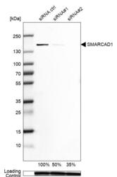
- Experimental details
- Western blot analysis in Rh30 cells transfected with control siRNA, target specific siRNA probe #1 and #2, using Anti-SMARCAD1 antibody. Remaining relative intensity is presented. Loading control: Anti-GAPDH.
- Sample type
- Human
- Protocol
- Protocol

Enhanced validation
Supportive validation
- Submitted by
- 55af80e3e0991
- Enhanced method
- Genetic validation
- Main image
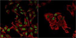
- Experimental details
- Confocal images of immunofluorescently stained human U-2 OS cells.The protein SMARCAD1 is shown in green and the microtubules in red. The image to the left show cells transfected with control siRNA and the image to the right show cells where SMARCAD1 has been downregulated with specific siRNA.
- Sample type
- U-2 OS cells
- Primary Ab dilution
- 1:200
- Secondary Ab
- Secondary Ab
- Secondary Ab dilution
- 1:800
- Knockdown/Genetic Approaches Application
- Immunocytochemistry

Supportive validation
- Submitted by
- Atlas Antibodies (provider)
- Main image
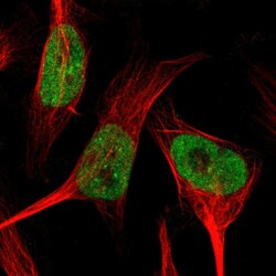
- Experimental details
- Immunofluorescent staining of human cell line U-251 MG shows localization to nucleoplasm.
- Sample type
- Human
