 Explore
 Validate
 Learn
Learn Immunocytochemistry
Antibody data
- Antibody Data
- Antigen structure
- References [2]
- Comments [0]
- Validations
- Immunocytochemistry [1]
- Flow cytometry [1]
Submit
Validation data
Reference
Comment
Report error
- Product number
- MAB1415 - Provider product page

- Provider
- R&D Systems
- Product name
- Human Glut3 Antibody
- Antibody type
- Monoclonal
- Description
- Protein A or G purified from hybridoma culture supernatant. Detects human Glut3. Recognizes human Glut3 expression on human Glut3-transfected NS0 cells, but not the NS0 control transfectants. No cross-reactivity was observed with transfectants expressing human Glut1 or human Glut2.
- Reactivity
- Human
- Host
- Mouse
- Conjugate
- Unconjugated
- Antigen sequence
P11169- Isotype
- IgG
- Antibody clone number
- 202017
- Vial size
- 200 ug
- Concentration
- LYOPH
- Storage
- Use a manual defrost freezer and avoid repeated freeze-thaw cycles. 12 months from date of receipt, -20 to -70 °C as supplied. 1 month, 2 to 8 °C under sterile conditions after reconstitution. 6 months, -20 to -70 °C under sterile conditions after reconstitution.
Submitted references Rhinovirus induces an anabolic reprogramming in host cell metabolism essential for viral replication.
IGF-I increases the recruitment of GLUT4 and GLUT3 glucose transporters on cell surface in hyperthyroidism.
Gualdoni GA, Mayer KA, Kapsch AM, Kreuzberg K, Puck A, Kienzl P, Oberndorfer F, Frühwirth K, Winkler S, Blaas D, Zlabinger GJ, Stöckl J
Proceedings of the National Academy of Sciences of the United States of America 2018 Jul 24;115(30):E7158-E7165
Proceedings of the National Academy of Sciences of the United States of America 2018 Jul 24;115(30):E7158-E7165
IGF-I increases the recruitment of GLUT4 and GLUT3 glucose transporters on cell surface in hyperthyroidism.
Dimitriadis G, Maratou E, Boutati E, Kollias A, Tsegka K, Alevizaki M, Peppa M, Raptis SA, Hadjidakis DJ
European journal of endocrinology 2008 Mar;158(3):361-6
European journal of endocrinology 2008 Mar;158(3):361-6
No comments: Submit comment
Supportive validation
- Submitted by
- R&D Systems (provider)
- Main image
- Experimental details
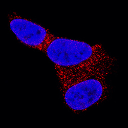
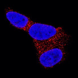
- Glut3 in SH-SY5Y Human Cell Line. Glut3 was detected in immersion fixed SH-SY5Y human neuroblastoma cell line using Mouse Anti-Human Glut3 Monoclonal Antibody (Catalog # MAB1415) at 25 µg/mL for 3 hours at room temperature. Cells were stained using the NorthernLights™ 557-conjugated Anti-Mouse IgG Secondary Antibody (red; Catalog # NL007) and counterstained with DAPI (blue). Specific staining was localized to cytoplasm. View our protocol for Fluorescent ICC Staining of Cells on Coverslips.
Supportive validation
- Submitted by
- R&D Systems (provider)
- Main image
- Experimental details
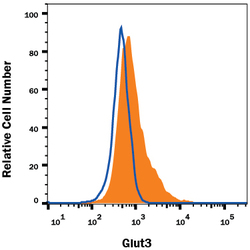
- Detection of Glut3 in NS0 Mouse Cell Line Transfected with Human Glut3 by Flow Cytometry. NS0 mouse myeloma cell line transfected with human Glut3 was stained with Mouse Anti-Human Glut3 Monoclonal Antibody (Catalog # MAB1415, filled histogram) or isotype control antibody (Catalog # MAB0041, open histogram), followed by Phycoerythrin-conjugated Anti-Mouse IgG Secondary Antibody (Catalog # F0102B). View our protocol for Staining Membrane-associated Proteins.
