 Explore
 Validate
 Learn
Learn Western blot
 ELISA
ELISAAntibody data
- Antibody Data
- Antigen structure
- References [37]
- Comments [0]
- Validations
- Western blot [1]
Submit
Validation data
Reference
Comment
Report error
- Product number
- 20570-1-AP - Provider product page

- Provider
- Proteintech Group
- Product name
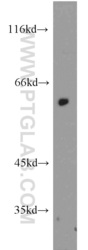
- PAX7 antibody
- Antibody type
- Polyclonal
- Description
- PAX7 antibody (Cat. #20570-1-AP) is a rabbit polyclonal antibody that shows reactivity with human, mouse and has been validated for the following applications: IF, WB,ELISA.
- Reactivity
- Human, Mouse
- Host
- Rabbit
- Conjugate
- Unconjugated
- Isotype
- IgG
- Vial size
- 20ul, 150ul
Submitted references Collagen-Based 3D Scaffolds from Sea Urchin Food Waste for Skeletal Muscle Tissue Engineering.
Lactiplantibacillus plantarum LM1001 Supplementation Attenuates Muscle Atrophy and Function Decline in Aged Mice.
Integrative Transcriptomic and Proteomic Analysis Reveals CaMK4-Mediated Regulation of Proliferation in Goat Skeletal Muscle Satellite Cells.
Neuromuscular electrical stimulation alleviates stroke-related sarcopenia by promoting satellite cells myogenic differentiation via AMPK-ULK1-Autophagy axis.
The Role of PAX7 in Breast Cancer Prognosis and Its Mechanistic Involvement in the Wnt/β-Catenin Pathway.
The mechanism of EGF in promoting skeletal muscle post-injury regeneration.
AAV-based TCAP delivery rescues mitochondria dislocation in limb-girdle muscular dystrophy R7.
The L-Ascorbic Acid Increases Proliferation and Differentiation of Yanbian Cattle Skeletal Muscle Satellite Cells by Activating the Akt/mTOR/P70S6K Signaling Pathway.
LMOD2 interaction with ACTC1 regulates myogenic differentiation.
The tumor suppressor pRb and its relative p130 are required to maintain murine adult skeletal muscle homeostasis.
CTCF Represses CIB2 to Balance Proliferation and Differentiation of Goat Myogenic Satellite Cells via Integrin α7β1-PI3K/AKT Axis.
Multi-omic integration of single-cell data uncovers methylation profiles of super-enhancers in skeletal muscle stem cells.
Tuina Promotes Repair of Chronic Cervical Muscle Injury by Regulating Satellite Cell Proliferation and Differentiation and Inhibiting Myocyte Apoptosis.
Inhibitor of Myom3 inhibits proliferation and promotes differentiation of sheep myoblasts.
The role and mechanism of β-catenin-mediated skeletal muscle satellite cells in osteoporotic fractures by Jian-Pi-Bu-Shen formula.
Irisin Ameliorates Muscle Atrophy by Inhibiting the Upregulation of the Ubiquitin‒Proteasome System in Chronic Kidney Disease.
Myogenic exosome miR-140-5p modulates skeletal muscle regeneration and injury repair by regulating muscle satellite cells.
Myoblast-derived ADAMTS-like 2 promotes skeletal muscle regeneration after injury.
Role and Regulatory Mechanism of circRNA_14820 in the Proliferation and Differentiation of Goat Skeletal Muscle Satellite Cells.
Therapeutic Myogenesis Induced by Ultrasound Exposure in a Volumetric Skeletal Muscle Loss Injury Model.
Urotensin II can Induce Skeletal Muscle Atrophy Associated with Upregulating Ubiquitin-Proteasome System and Inhibiting the Differentiation of Satellite Cells in CRF Mice.
A Novel in Duck Myoblasts: The Transcription Factor Retinoid X Receptor Alpha (RXRA) Inhibits Lipid Accumulation by Promoting CD36 Expression.
Apolipoprotein H: a novel regulator of fat accumulation in duck myoblasts.
Role of lncRNA Has2os in Skeletal Muscle Differentiation and Regeneration.
Time-Series Clustering of lncRNA-mRNA Expression during the Adipogenic Transdifferentiation of Porcine Skeletal Muscle Satellite Cells.
TGM2 positively regulates myoblast differentiation via enhancing the mTOR signaling.
circRNAome profiling reveals circFgfr2 regulates myogenesis and muscle regeneration via a feedback loop.
Myoblast-derived exosomes promote the repair and regeneration of injured skeletal muscle in mice.
CircUBE2Q2 promotes differentiation of cattle muscle stem cells and is a potential regulatory molecule of skeletal muscle development.
PERK Signaling Controls Myoblast Differentiation by Regulating MicroRNA Networks.
Trio cooperates with Myh9 to regulate neural crest-derived craniofacial development.
Extracellular vesicles derived from oesophageal cancer containing P4HB promote muscle wasting via regulating PHGDH/Bcl-2/caspase-3 pathway.
Overexpression of Dnmt3a ameliorates diabetic muscle atrophy by modulating the Pten/Akt pathway.
Exosomes of oral squamous cell carcinoma cells containing miR-181a-3p induce muscle cell atrophy and apoptosis by transmissible endoplasmic reticulum stress signaling.
KLF2 in Myeloid Lineage Cells Regulates the Innate Immune Response during Skeletal Muscle Injury and Regeneration.
Dilated cardiomyopathy-mediated heart failure induces a unique skeletal muscle myopathy with inflammation.
Anti-aging Effect of Transplanted Amniotic Membrane Mesenchymal Stem Cells in a Premature Aging Model of Bmi-1 Deficiency.
Akyürek EE, Melotti L, Erba M, Carolo A, Martinelli G, Roncoroni M, Marzorati S, Patruno M, Sugni M, Sacchetto R
Animals : an open access journal from MDPI 2026 Feb 5;16(3)
Animals : an open access journal from MDPI 2026 Feb 5;16(3)
Lactiplantibacillus plantarum LM1001 Supplementation Attenuates Muscle Atrophy and Function Decline in Aged Mice.
Karekezi J, Kim H, Dusabimana T, Nugroho TA, Ndahigwa EN, So YJ, Kim J, Kim TR, Sohn M, Miao J, Moon Y, Park SW
Nutrients 2025 Oct 4;17(19)
Nutrients 2025 Oct 4;17(19)
Integrative Transcriptomic and Proteomic Analysis Reveals CaMK4-Mediated Regulation of Proliferation in Goat Skeletal Muscle Satellite Cells.
Cong H, Xu L, Liu Y, Wang Z, Ren T, Ruan P, Zhang H, Liu C, Han Y, Hu P, Zeng Y, Ceccobelli S, E G
Animals : an open access journal from MDPI 2025 Oct 24;15(21)
Animals : an open access journal from MDPI 2025 Oct 24;15(21)
Neuromuscular electrical stimulation alleviates stroke-related sarcopenia by promoting satellite cells myogenic differentiation via AMPK-ULK1-Autophagy axis.
Xiang X, Huang L, Luo W, Qin L, Bian M, Chen W, Han G, Wang N, Mo G, Zhang C, Zhang Y, Yang H, Lu S, Zhang J, Fu T
Journal of orthopaedic translation 2025 May;52:249-264
Journal of orthopaedic translation 2025 May;52:249-264
The Role of PAX7 in Breast Cancer Prognosis and Its Mechanistic Involvement in the Wnt/β-Catenin Pathway.
Ge Q, Zhang W, Li C, Li X, Wang Z, Li X
Journal of cellular and molecular medicine 2025 May;29(10):e70602
Journal of cellular and molecular medicine 2025 May;29(10):e70602
The mechanism of EGF in promoting skeletal muscle post-injury regeneration.
Teng H, Liu Y, Hao R, Zhang L, Zhang X, Li S, Li S, Tong H
Differentiation; research in biological diversity 2025 May-Jun;143:100862
Differentiation; research in biological diversity 2025 May-Jun;143:100862
AAV-based TCAP delivery rescues mitochondria dislocation in limb-girdle muscular dystrophy R7.
Lv X, Liu S, Li X, Lv H, Shao K, Luo S, Zhao D, Yan C, Lin P
Brain : a journal of neurology 2025 May 13;148(5):1680-1694
Brain : a journal of neurology 2025 May 13;148(5):1680-1694
The L-Ascorbic Acid Increases Proliferation and Differentiation of Yanbian Cattle Skeletal Muscle Satellite Cells by Activating the Akt/mTOR/P70S6K Signaling Pathway.
Jin H, Li Q, Tang L, Naseem S, Park S, Wang E, Sun B, Manzoor A, Hur SJ, Li X, Choi SH
Food science of animal resources 2025 Mar;45(2):484-503
Food science of animal resources 2025 Mar;45(2):484-503
LMOD2 interaction with ACTC1 regulates myogenic differentiation.
Wang K, Liu C, Yi L, Liufu S, Chen W, Liu X, Chen B, Xu X, Liu J, Liu X, Yin Y, Ma H
BMC genomics 2025 Jul 31;26(1):709
BMC genomics 2025 Jul 31;26(1):709
The tumor suppressor pRb and its relative p130 are required to maintain murine adult skeletal muscle homeostasis.
Jiang Z, Baechler BL, Li H, Lad H, Ciavarra G, Ben-David Y, Burden SJ, Quadrilatero J, Zacksenhaus E
Oncogene 2025 Aug;44(30):2662-2674
Oncogene 2025 Aug;44(30):2662-2674
CTCF Represses CIB2 to Balance Proliferation and Differentiation of Goat Myogenic Satellite Cells via Integrin α7β1-PI3K/AKT Axis.
Gong C, Song H, Hao Z, Zhang Z, Luo N, Chen X
Cells 2025 Aug 5;14(15)
Cells 2025 Aug 5;14(15)
Multi-omic integration of single-cell data uncovers methylation profiles of super-enhancers in skeletal muscle stem cells.
Zeng A, Liu H, He S, Luo X, Zhang Z, Fu M, Yu B
Epigenetics & chromatin 2025 Aug 11;18(1):54
Epigenetics & chromatin 2025 Aug 11;18(1):54
Tuina Promotes Repair of Chronic Cervical Muscle Injury by Regulating Satellite Cell Proliferation and Differentiation and Inhibiting Myocyte Apoptosis.
Zhang J, Qu S, Huang Y, Zhang X, Tong X, Fang Y, Rao T, Liu K, Lin J, Lin Y, Zeng C, Zhang G, Jing X, Liao J, Kan Y
Journal of pain research 2024;17:3419-3429
Journal of pain research 2024;17:3419-3429
Inhibitor of Myom3 inhibits proliferation and promotes differentiation of sheep myoblasts.
Kong L, Yuan C, Guo T, Sun L, Liu J, Lu Z
Genomics 2024 Sep;116(5):110921
Genomics 2024 Sep;116(5):110921
The role and mechanism of β-catenin-mediated skeletal muscle satellite cells in osteoporotic fractures by Jian-Pi-Bu-Shen formula.
Tang Y, Mu Z, Pan D, Liu R, Hong S, Xiong Z
Journal of molecular histology 2024 Oct;55(5):875-893
Journal of molecular histology 2024 Oct;55(5):875-893
Irisin Ameliorates Muscle Atrophy by Inhibiting the Upregulation of the Ubiquitin‒Proteasome System in Chronic Kidney Disease.
Wang S, Pan Y, Pang Q, Zhang A
Calcified tissue international 2024 Nov;115(5):712-724
Calcified tissue international 2024 Nov;115(5):712-724
Myogenic exosome miR-140-5p modulates skeletal muscle regeneration and injury repair by regulating muscle satellite cells.
Cao X, Xue L, Yu X, Yan Y, Lu J, Luo X, Wang H, Wang J
Aging 2024 Feb 29;16(5):4609-4630
Aging 2024 Feb 29;16(5):4609-4630
Myoblast-derived ADAMTS-like 2 promotes skeletal muscle regeneration after injury.
Taye N, Rodriguez L, Iatridis JC, Han WM, Hubmacher D
NPJ Regenerative medicine 2024 Dec 19;9(1):39
NPJ Regenerative medicine 2024 Dec 19;9(1):39
Role and Regulatory Mechanism of circRNA_14820 in the Proliferation and Differentiation of Goat Skeletal Muscle Satellite Cells.
Yang P, Li X, Liu C, Han Y, E G, Huang Y
International journal of molecular sciences 2024 Aug 15;25(16)
International journal of molecular sciences 2024 Aug 15;25(16)
Therapeutic Myogenesis Induced by Ultrasound Exposure in a Volumetric Skeletal Muscle Loss Injury Model.
Mohamad Yusoff F, Nakashima A, Kajikawa M, Kishimoto S, Maruhashi T, Higashi Y
The American journal of sports medicine 2023 Nov;51(13):3554-3566
The American journal of sports medicine 2023 Nov;51(13):3554-3566
Urotensin II can Induce Skeletal Muscle Atrophy Associated with Upregulating Ubiquitin-Proteasome System and Inhibiting the Differentiation of Satellite Cells in CRF Mice.
Pan Y, Zhou T, Dong X, Wu L, Wang P, Wang S, Zhang A
Calcified tissue international 2023 May;112(5):603-612
Calcified tissue international 2023 May;112(5):603-612
A Novel in Duck Myoblasts: The Transcription Factor Retinoid X Receptor Alpha (RXRA) Inhibits Lipid Accumulation by Promoting CD36 Expression.
Pan Z, Chen X, Wu D, Li X, Gao W, Li G, Du G, Zhang C, Jin S, Geng Z
International journal of molecular sciences 2023 Jan 7;24(2)
International journal of molecular sciences 2023 Jan 7;24(2)
Apolipoprotein H: a novel regulator of fat accumulation in duck myoblasts.
Pan Z, Du G, Li G, Wu D, Chen X, Geng Z
Journal of animal science and technology 2022 Nov;64(6):1199-1214
Journal of animal science and technology 2022 Nov;64(6):1199-1214
Role of lncRNA Has2os in Skeletal Muscle Differentiation and Regeneration.
Chen W, Chen W, Liu P, Qian S, Tao S, Huang M, Xu W, Li C, Chen X, Lin H, Qin Z, Lu J, Xie S
Cells 2022 Nov 4;11(21)
Cells 2022 Nov 4;11(21)
Time-Series Clustering of lncRNA-mRNA Expression during the Adipogenic Transdifferentiation of Porcine Skeletal Muscle Satellite Cells.
Qiu X, Gao G, Du L, Wang J, Wang Q, Yang F, Zhou X, Long D, Huang J, Liu Z, Qi R
Current issues in molecular biology 2022 May 6;44(5):2038-2053
Current issues in molecular biology 2022 May 6;44(5):2038-2053
TGM2 positively regulates myoblast differentiation via enhancing the mTOR signaling.
Wang D, Zhao D, Li Y, Dai T, Liu F, Yan C
Biochimica et biophysica acta. Molecular cell research 2022 Mar;1869(3):119173
Biochimica et biophysica acta. Molecular cell research 2022 Mar;1869(3):119173
circRNAome profiling reveals circFgfr2 regulates myogenesis and muscle regeneration via a feedback loop.
Yan J, Yang Y, Fan X, Liang G, Wang Z, Li J, Wang L, Chen Y, Adetula AA, Tang Y, Li K, Wang D, Tang Z
Journal of cachexia, sarcopenia and muscle 2022 Feb;13(1):696-712
Journal of cachexia, sarcopenia and muscle 2022 Feb;13(1):696-712
Myoblast-derived exosomes promote the repair and regeneration of injured skeletal muscle in mice.
Ji S, Ma P, Cao X, Wang J, Yu X, Luo X, Lu J, Hou W, Zhang Z, Yan Y, Dong Y, Wang H
FEBS open bio 2022 Dec;12(12):2213-2226
FEBS open bio 2022 Dec;12(12):2213-2226
CircUBE2Q2 promotes differentiation of cattle muscle stem cells and is a potential regulatory molecule of skeletal muscle development.
Zhang RM, Pan Y, Zou CX, An Q, Cheng JR, Li PJ, Zheng ZH, Pan Y, Feng WY, Yang SF, Shi DS, Wei YM, Deng YF
BMC genomics 2022 Apr 6;23(1):267
BMC genomics 2022 Apr 6;23(1):267
PERK Signaling Controls Myoblast Differentiation by Regulating MicroRNA Networks.
Tan YY, Zhang Y, Li B, Ou YW, Xie SJ, Chen PP, Mei SQ, Huang QJ, Zheng LL, Qu LH
Frontiers in cell and developmental biology 2021;9:670435
Frontiers in cell and developmental biology 2021;9:670435
Trio cooperates with Myh9 to regulate neural crest-derived craniofacial development.
Guo S, Meng L, Liu H, Yuan L, Zhao N, Ni J, Zhang Y, Ben J, Li YP, Ma J
Theranostics 2021;11(9):4316-4334
Theranostics 2021;11(9):4316-4334
Extracellular vesicles derived from oesophageal cancer containing P4HB promote muscle wasting via regulating PHGDH/Bcl-2/caspase-3 pathway.
Gao X, Wang Y, Lu F, Chen X, Yang D, Cao Y, Zhang W, Chen J, Zheng L, Wang G, Fu M, Ma L, Song Y, Zhan Q
Journal of extracellular vesicles 2021 Mar;10(5):e12060
Journal of extracellular vesicles 2021 Mar;10(5):e12060
Overexpression of Dnmt3a ameliorates diabetic muscle atrophy by modulating the Pten/Akt pathway.
Wang M, Wu X, Gan L, Teng Z, Zhang H, Zhang Y
Experimental physiology 2020 Nov;105(11):1918-1927
Experimental physiology 2020 Nov;105(11):1918-1927
Exosomes of oral squamous cell carcinoma cells containing miR-181a-3p induce muscle cell atrophy and apoptosis by transmissible endoplasmic reticulum stress signaling.
Qiu L, Chen W, Wu C, Yuan Y, Li Y
Biochemical and biophysical research communications 2020 Dec 17;533(4):831-837
Biochemical and biophysical research communications 2020 Dec 17;533(4):831-837
KLF2 in Myeloid Lineage Cells Regulates the Innate Immune Response during Skeletal Muscle Injury and Regeneration.
Manoharan P, Song T, Radzyukevich TL, Sadayappan S, Lingrel JB, Heiny JA
iScience 2019 Jul 26;17:334-346
iScience 2019 Jul 26;17:334-346
Dilated cardiomyopathy-mediated heart failure induces a unique skeletal muscle myopathy with inflammation.
Song T, Manoharan P, Millay DP, Koch SE, Rubinstein J, Heiny JA, Sadayappan S
Skeletal muscle 2019 Jan 24;9(1):4
Skeletal muscle 2019 Jan 24;9(1):4
Anti-aging Effect of Transplanted Amniotic Membrane Mesenchymal Stem Cells in a Premature Aging Model of Bmi-1 Deficiency.
Xie C, Jin J, Lv X, Tao J, Wang R, Miao D
Scientific reports 2015 Sep 15;5:13975
Scientific reports 2015 Sep 15;5:13975
No comments: Submit comment
Supportive validation
- Submitted by
- Proteintech Group (provider)
- Main image
- Experimental details
- The PAX7 antibody from Proteintech is a rabbit polyclonal antibody to a peptide of human PAX7. This antibody recognizes human antigen. The PAX7 antibody has been validated for the following applications: ELISA, WB analysis.
