Explore
 Validate
 Learn
Learn Western blot
Antibody data
- Antibody Data
- Antigen structure
- References [0]
- Comments [0]
- Validations
- Western blot [2]
- Immunocytochemistry [3]
- Immunoprecipitation [1]
Submit
Validation data
Reference
Comment
Report error
- Product number
- GTX54533 - Provider product page

- Provider
- GeneTex
- Product name
- PAX7 antibody
- Antibody type
- Polyclonal
- Reactivity
- Human, Mouse
- Host
- Rabbit
No comments: Submit comment
Supportive validation
- Submitted by
- GeneTex (provider)
- Main image

- Experimental details
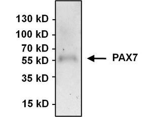
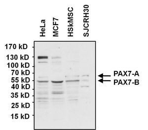
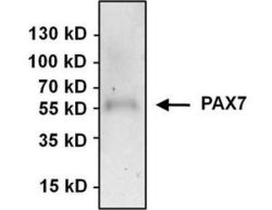
- WB analysis of various cell lysates (50 ug per lane) using PAX7 antibody at a dilution of 1:1000.
- Submitted by
- GeneTex (provider)
- Main image

- Experimental details
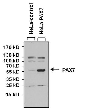
- WB analysis of lysates from HeLa cells overexpressing PAX7 (right) or empty vector control (left) using PAX7 antibody at a dilution of 1:1000.
Supportive validation
- Submitted by
- GeneTex (provider)
- Main image

- Experimental details
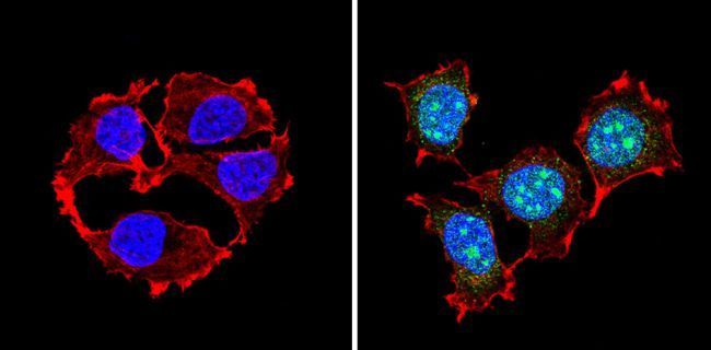
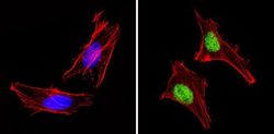
- ICC/IF analysis of MCF7 cells with (right) or without (left) PAX7 antibody at a dilution of 1:100 (green). F-actin (red) was stained with a flourescent red phalloidin and nuclei (blue) were stained with Hoechst or DAPI. Images were taken at a magnification of 60x.
- Submitted by
- GeneTex (provider)
- Main image
- Experimental details
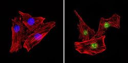
- ICC/IF analysis of HeLa cells with (right) or without (left) PAX7 antibody at a dilution of 1:100 (green). F-actin (red) was stained with a flourescent red phalloidin and nuclei (blue) were stained with Hoechst or DAPI. Images were taken at a magnification of 60x.

- Submitted by
- GeneTex (provider)
- Main image
- Experimental details
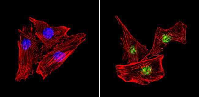
- ICC/IF analysis of C2C12 cells with (right) or without (left) PAX7 antibody at a dilution of 1:100 (green). F-actin (red) was stained with a flourescent red phalloidin and nuclei (blue) were stained with Hoechst or DAPI. Images were taken at a magnification of 60x.
Supportive validation
- Submitted by
- GeneTex (provider)
- Main image
- Experimental details
- IP analysis was performed using 500 ug of HeLa lysate and 3 ug of PAX7 antibody. The precipitate was detected by the same antibody with EasyBlot as the secondary antibody.