Explore
 Validate
 Learn
Learn Western blot
 Immunocytochemistry
Antibody data
- Antibody Data
- Antigen structure
- References [3]
- Comments [0]
- Validations
- Immunocytochemistry [4]
- Other assay [3]
Submit
Validation data
Reference
Comment
Report error
- Product number
- PA5-68506 - Provider product page

- Provider
- Invitrogen Antibodies
- Product name
- PAX7 Polyclonal Antibody
- Antibody type
- Polyclonal
- Antigen
- Synthetic peptide
- Description
- This target displays homology in the following species: Cow: 100%; Dog: 100%; Goat: 86%; Guinea Pig: 100%; Human: 100%; Mouse: 100%; Rabbit: 100%; Rat: 100%; Zebrafish: 100%
- Reactivity
- Human, Mouse, Rat
- Host
- Rabbit
- Isotype
- IgG
- Vial size
- 100 μL
- Concentration
- 0.5 mg/mL
- Storage
- -20°C, Avoid Freeze/Thaw Cycles
Submitted references Simple and effective serum-free medium for sustained expansion of bovine satellite cells for cell cultured meat.
Effects of Hypoxia on Proliferation and Differentiation in Belgian Blue and Hanwoo Muscle Satellite Cells for the Development of Cultured Meat.
Extracellular Heme Proteins Influence Bovine Myosatellite Cell Proliferation and the Color of Cell-Based Meat.
Stout AJ, Mirliani AB, Rittenberg ML, Shub M, White EC, Yuen JSK Jr, Kaplan DL
Communications biology 2022 Jun 2;5(1):466
Communications biology 2022 Jun 2;5(1):466
Effects of Hypoxia on Proliferation and Differentiation in Belgian Blue and Hanwoo Muscle Satellite Cells for the Development of Cultured Meat.
Park S, Gagliardi M, Swennen G, Dogan A, Kim Y, Park Y, Park G, Oh S, Post M, Choi J
Biomolecules 2022 Jun 16;12(6)
Biomolecules 2022 Jun 16;12(6)
Extracellular Heme Proteins Influence Bovine Myosatellite Cell Proliferation and the Color of Cell-Based Meat.
Simsa R, Yuen J, Stout A, Rubio N, Fogelstrand P, Kaplan DL
Foods (Basel, Switzerland) 2019 Oct 21;8(10)
Foods (Basel, Switzerland) 2019 Oct 21;8(10)
No comments: Submit comment
Supportive validation
- Submitted by
- Invitrogen Antibodies (provider)
- Main image
- Experimental details
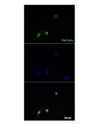
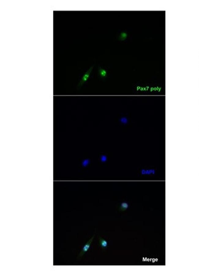
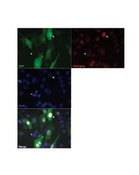
- Immunofluorescent analysis of PAX7 in C2C12 cells using PAX7 polyclonal antibody (Product # PA5-68506).

- Submitted by
- Invitrogen Antibodies (provider)
- Main image
- Experimental details
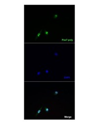
- Immunofluorescent analysis of PAX7 in C2C12 cells using PAX7 polyclonal antibody (Product # PA5-68506).
- Submitted by
- Invitrogen Antibodies (provider)
- Main image
- Experimental details
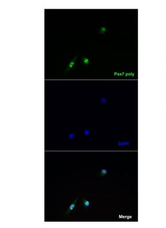
- Immunofluorescent analysis of PAX7 in C2C12 cells using PAX7 polyclonal antibody (Product # PA5-68506).

- Submitted by
- Invitrogen Antibodies (provider)
- Main image
- Experimental details
- Immunofluorescent analysis of PAX7 in C2C12 cells using PAX7 polyclonal antibody (Product # PA5-68506).
Supportive validation
- Submitted by
- Invitrogen Antibodies (provider)
- Main image
- Experimental details
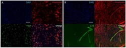
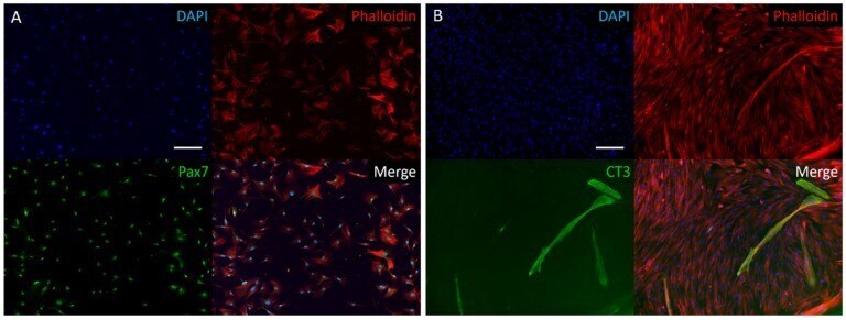
- Figure 1 Two-dimensional immunofluorescence stain of isolated bovine muscle satellite cells (BSCs). ( A ) Proliferating bovine satellite stained for DAPI, actin cytoskeleton (Phalloidin), and Pax7, a nuclear marker of satellite cells. Stains show a highly pure satellite cell population, following isolation and pre-plating protocol. ( B ) Following one week of differentiation, cells were stained for DAPI, actin cytoskeleton (Phalloidin), and Troponin T (CT3), a marker of myogenesis. Scale bars are 200 um.
- Submitted by
- Invitrogen Antibodies (provider)
- Main image
- Experimental details
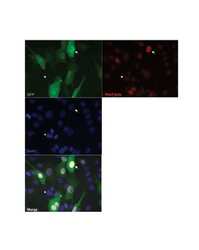
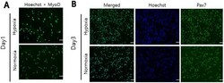
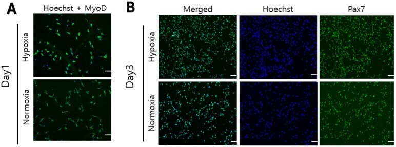
- The cell nuclei stained with Hoechst (Hoechst 33,342 nucleic acid stain) were blue fluorescence and Pax7 and MyoD proteins were stained with green fluorescence. ( A ): The nuclei and MyoD protein of Hanwoo myosatellite cells cultured for 1 day in hypoxia (2% O 2 ) and normoxia (20% O 2 ) were stained. ( B , C ): The nuclei, Pax7 and MyoD protein of Hanwoo myosatellite cells cultured for 3 days in hypoxia (2% O 2 ) and normoxia (20% O 2 ) were stained. Experiments were performed in triplicate and repeated three times (* p < 0.05). Hanwoo myosatellite cells were seeded in T25 flasks at 1800 cells/cm 2 and cultured in GM for 6 days in normoxia (20% O 2 ) or hypoxia (2% O 2 ) ( D ). ( E ): Relative Pax7, Myf5, MyoD and HIF1alpha mRNA levels were compared in Hanwoo myosatellite cells from ( D ). GAPDH was used as an internal control for RT-PCR. scale bar: 100 mum.
- Submitted by
- Invitrogen Antibodies (provider)
- Main image
- Experimental details
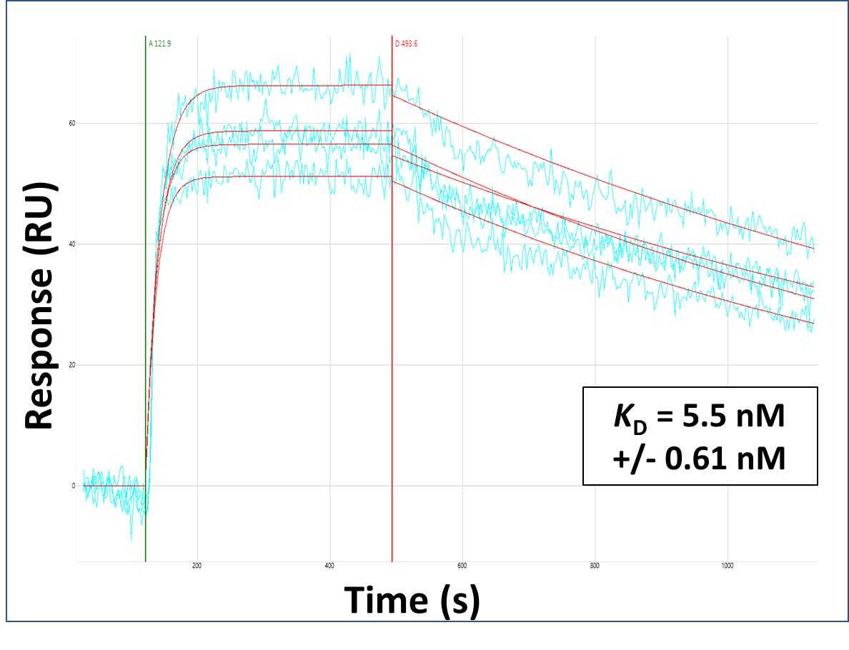
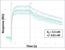
- Surface Plasmon Resonance of PAX7 polyclonal antibody (Product # PA5-68506). Purified polyclonal antibodies were immobilized on a Protein A/G coated Carterra LSA sensor chip at concentrations of 5, and 50 µg/mL in duplicate. Antibodies on the surface were exposed to interaction with peptides sequentially via microfluidic controlled flow at 333 nm peptide concentration for kinetic characterization of the binders for affinity and specificity, followed by curve fitting using the Kinetics software. Kd determinations for both concentrations were averaged and results and standard deviation are shown.