 Explore
 Validate
 Learn
Learn Western blot
 Immunohistochemistry
Antibody data
- Antibody Data
- Antigen structure
- References [4]
- Comments [0]
- Validations
- Western blot [1]
- Immunohistochemistry [1]
Submit
Validation data
Reference
Comment
Report error
- Product number
- AMAb91124 - Provider product page

- Provider
- Atlas Antibodies
- Proper citation
- Atlas Antibodies Cat#AMAb91124, RRID:AB_2665809
- Product name
- Anti-LAMA5
- Antibody type
- Monoclonal
- Description
- Monoclonal Antibody against Human LAMA5, Clone ID: CL3118, Gene description: Laminin subunit alpha 5, Validated applications: WB, IHC, Uniprot ID: O15230, Storage: Store at +4°C for short term storage. Long time storage is recommended at -20°C.
- Reactivity
- Human
- Host
- Mouse
- Conjugate
- Unconjugated
- Isotype
- IgG
- Antibody clone number
- CL3118
- Vial size
- 100 µl
- Concentration
- 1.0 mg/ml
- Storage
- Store at +4°C for short term storage. Long time storage is recommended at -20°C.
- Handling
- The antibody solution should be gently mixed before use.
Submitted references Basal cell adhesion molecule promotes metastasis-associated processes in ovarian cancer.
Limbal BCAM expression identifies a proliferative progenitor population capable of holoclone formation and corneal differentiation.
Clear Evidence of LAMA5 Gene Biallelic Truncating Variants Causing Infantile Nephrotic Syndrome.
Parallel Murine and Human Plaque Proteomics Reveals Pathways of Plaque Rupture.
Sivakumar S, Lieber S, Librizzi D, Keber C, Sommerfeld L, Finkernagel F, Roth K, Reinartz S, Bartsch JW, Graumann J, Müller-Brüsselbach S, Müller R
Clinical and translational medicine 2023 Jan;13(1):e1176
Clinical and translational medicine 2023 Jan;13(1):e1176
Limbal BCAM expression identifies a proliferative progenitor population capable of holoclone formation and corneal differentiation.
Sasamoto Y, Lee CAA, Wilson BJ, Buerger F, Martin G, Mishra A, Kiritoshi S, Tran J, Gonzalez G, Hildebrandt F, Jo VY, Lian CG, Murphy GF, Ksander BR, Frank MH, Frank NY
Cell reports 2022 Aug 9;40(6):111166
Cell reports 2022 Aug 9;40(6):111166
Clear Evidence of LAMA5 Gene Biallelic Truncating Variants Causing Infantile Nephrotic Syndrome.
Taniguchi Y, Nagano C, Sekiguchi K, Tashiro A, Sugawara N, Sakaguchi H, Umeda C, Aoto Y, Ishiko S, Rossanti R, Sakakibara N, Horinouchi T, Yamamura T, Kondo A, Nagai S, Nagase H, Iijima K, Miner JH, Nozu K
Kidney360 2021 Dec 30;2(12):1968-1978
Kidney360 2021 Dec 30;2(12):1968-1978
Parallel Murine and Human Plaque Proteomics Reveals Pathways of Plaque Rupture.
Vaisar T, Hu JH, Airhart N, Fox K, Heinecke J, Nicosia RF, Kohler T, Potter ZE, Simon GM, Dix MM, Cravatt BF, Gharib SA, Dichek DA
Circulation research 2020 Sep 25;127(8):997-1022
Circulation research 2020 Sep 25;127(8):997-1022
No comments: Submit comment
Enhanced validation
- Submitted by
- Atlas Antibodies (provider)
- Enhanced method
- Genetic validation
- Main image

- Experimental details
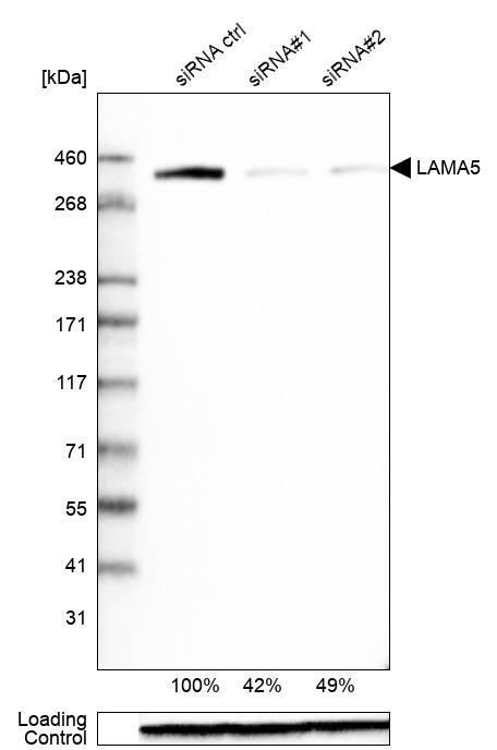
- Western blot analysis in Caco-2 cells transfected with control siRNA, target specific siRNA probe #1 and #2, using Anti-LAMA5 antibody. Remaining relative intensity is presented. Loading control: Anti-GAPDH.
- Sample type
- Human
- Protocol
- Protocol
Supportive validation
- Submitted by
- Atlas Antibodies (provider)
- Enhanced method
- Orthogonal validation
- Main image
- Experimental details
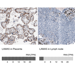
- Immunohistochemistry analysis in human placenta and lymph node tissues using AMAb91124 antibody. Corresponding LAMA5 RNA-seq data are presented for the same tissues.
- Sample type
- Human
- Protocol
- Protocol
