 Explore
 Validate
 Learn
Learn Western blot
 Immunocytochemistry
Antibody data
- Antibody Data
- Antigen structure
- References [2]
- Comments [0]
- Validations
- Western blot [1]
- Immunocytochemistry [1]
- Immunohistochemistry [1]
Submit
Validation data
Reference
Comment
Report error
- Product number
- HPA029725 - Provider product page

- Provider
- Atlas Antibodies
- Proper citation
- Atlas Antibodies Cat#HPA029725, RRID:AB_10602328
- Product name
- Anti-ASRGL1
- Antibody type
- Polyclonal
- Description
- Polyclonal Antibody against Human ASRGL1, Gene description: asparaginase like 1, Alternative Gene Names: ALP, ALP1, FLJ22316, Validated applications: ICC, IHC, WB, Uniprot ID: Q7L266, Storage: Store at +4°C for short term storage. Long time storage is recommended at -20°C.
- Reactivity
- Human
- Host
- Rabbit
- Conjugate
- Unconjugated
- Isotype
- IgG
- Vial size
- 100 µl
- Concentration
- 0.1 mg/ml
- Storage
- Store at +4°C for short term storage. Long time storage is recommended at -20°C.
- Handling
- The antibody solution should be gently mixed before use.
Submitted references TDP-43 proteinopathy in ALS is triggered by loss of ASRGL1 and associated with HML-2 expression.
Loss of ASRGL1 expression is an independent biomarker for disease-specific survival in endometrioid endometrial carcinoma.
Garcia-Montojo M, Fathi S, Rastegar C, Simula ER, Doucet-O'Hare T, Cheng YHH, Abrams RPM, Pasternack N, Malik N, Bachani M, Disanza B, Maric D, Lee MH, Wang H, Santamaria U, Li W, Sampson K, Lorenzo JR, Sanchez IE, Mezghrani A, Li Y, Sechi LA, Pineda S, Heiman M, Kellis M, Steiner J, Nath A
Nature communications 2024 May 16;15(1):4163
Nature communications 2024 May 16;15(1):4163
Loss of ASRGL1 expression is an independent biomarker for disease-specific survival in endometrioid endometrial carcinoma.
Edqvist PH, Huvila J, Forsström B, Talve L, Carpén O, Salvesen HB, Krakstad C, Grénman S, Johannesson H, Ljungqvist O, Uhlén M, Pontén F, Auranen A
Gynecologic oncology 2015 Jun;137(3):529-37
Gynecologic oncology 2015 Jun;137(3):529-37
No comments: Submit comment
Enhanced validation
- Submitted by
- Atlas Antibodies (provider)
- Enhanced method
- Recombinant expression validation
- Main image
- Experimental details
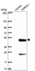
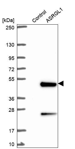
- Western blot analysis in control (vector only transfected HEK293T lysate) and ASRGL1 over-expression lysate (Co-expressed with a C-terminal myc-DDK tag (~3.1 kDa) in mammalian HEK293T cells, LY421255).
- Sample type
- Human
- Protocol
- Protocol
Supportive validation
- Submitted by
- Atlas Antibodies (provider)
- Main image

- Experimental details
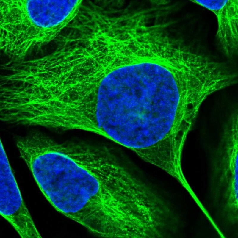
- Immunofluorescent staining of human cell line U-2 OS shows localization to nucleoplasm & microtubules.
- Sample type
- Human
Supportive validation
- Submitted by
- Atlas Antibodies (provider)
- Enhanced method
- Orthogonal validation
- Main image
- Experimental details
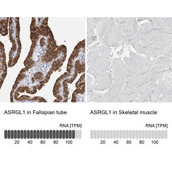
- Immunohistochemistry analysis in human fallopian tube and skeletal muscle tissues using HPA029725 antibody. Corresponding ASRGL1 RNA-seq data are presented for the same tissues.
- Sample type
- Human
- Protocol
- Protocol
