Explore
 Validate
 Learn
Learn Western blot
 Immunocytochemistry
 Immunohistochemistry
Antibody data
- Antibody Data
- Antigen structure
- References [0]
- Comments [0]
- Validations
- Immunocytochemistry [6]
- Immunohistochemistry [3]
Submit
Validation data
Reference
Comment
Report error
- Product number
- LS-C112408 - Provider product page

- Provider
- LSBio
- Product name
- AHNAK Antibody (clone EM-09) LS-C112408
- Antibody type
- Monoclonal
- Description
- Purified by protein-A affinity chromatography. Greater than 95% by SDS-PAGE.
- Reactivity
- Human, Mouse
- Host
- Mouse
- Isotype
- IgG
- Antibody clone number
- EM-09
- Storage
- Store at 2°C to 8°C. Do not freeze.
No comments: Submit comment
Supportive validation
- Submitted by
- LSBio (provider)
- Enhanced method
- Genetic validation
- Main image

- Experimental details
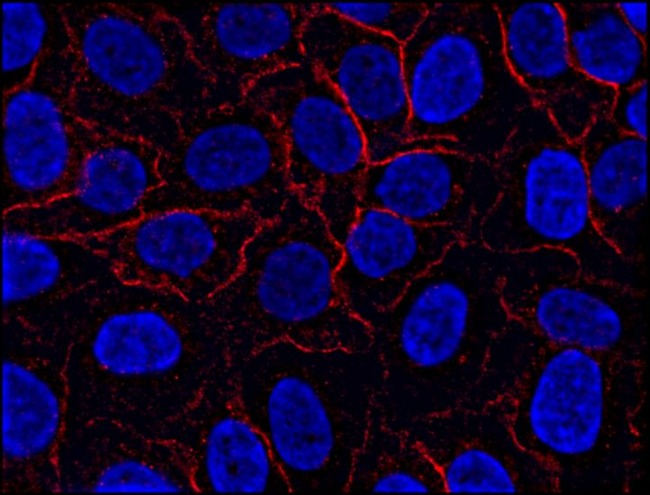
- Immunofluorescence staining of AHNAK1 in HeLa cell line using anti-AHNAK1 (EM-09; red). Cell nuclei stained with DAPI (blue).

- Submitted by
- LSBio (provider)
- Enhanced method
- Genetic validation
- Main image

- Experimental details
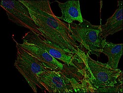
- Immunofluorescence staining of AHNAK1 in human primary fibroblasts using anti-AHNAK1 (EM-09; green). Actin filaments were decorated by phalloidin (red) and cell nuclei stained with DAPI (blue).

- Submitted by
- LSBio (provider)
- Main image
- Experimental details
- Immunofluorescence staining of AHNAK1 in human primary fibroblasts using anti-AHNAK1 (EM-09; green). Actin filaments were decorated by phalloidin (red) and cell nuclei stained with DAPI (blue).

- Submitted by
- LSBio (provider)
- Main image

- Experimental details
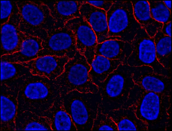
- Immunofluorescence staining of AHNAK1 in HeLa cell line using anti-AHNAK1 (EM-09; red). Cell nuclei stained with DAPI (blue).
- Submitted by
- LSBio (provider)
- Main image

- Experimental details
- Immunofluorescence staining of AHNAK1 in HeLa cell line using anti-AHNAK1 (EM-09; red). Cell nuclei stained with DAPI (blue).
- Submitted by
- LSBio (provider)
- Main image

- Experimental details
- Immunofluorescence staining of AHNAK1 in human primary fibroblasts using anti-AHNAK1 (EM-09; green). Actin filaments were decorated by phalloidin (red) and cell nuclei stained with DAPI (blue).

Enhanced validation
- Submitted by
- LSBio (provider)
- Enhanced method
- Genetic validation
- Main image

- Experimental details
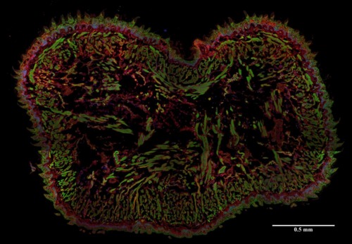
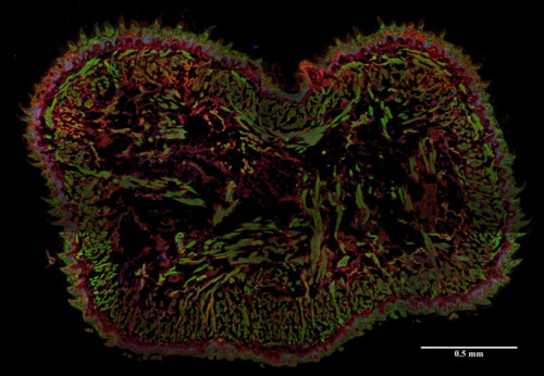
- Immunohistochemistry staining (frozen sections) of murine tongue by anti-AHNAK1 antibody (EM-09; red). Actin filaments were decorated by phalloidin (green), cell nuclei stained with DAPI (blue).
- Submitted by
- LSBio (provider)
- Enhanced method
- Genetic validation
- Main image

- Experimental details
- Immunohistochemistry staining (frozen sections) of murine tongue by anti-AHNAK1 antibody (EM-09; red). Actin filaments were decorated by phalloidin (green), cell nuclei stained with DAPI (blue).

- Submitted by
- LSBio (provider)
- Main image

- Experimental details
- Immunohistochemistry staining (frozen sections) of murine tongue by anti-AHNAK1 antibody (EM-09; red). Actin filaments were decorated by phalloidin (green), cell nuclei stained with DAPI (blue).