 Explore
 Validate
 Learn
Learn Western blot
Antibody data
- Antibody Data
- Antigen structure
- References [4]
- Comments [0]
- Validations
- Western blot [1]
- ELISA [1]
- Immunoprecipitation [1]
Submit
Validation data
Reference
Comment
Report error
- Product number
- H00079026-M01 - Provider product page

- Provider
- Abnova Corporation
- Proper citation
- Abnova Corporation Cat#H00079026-M01, RRID:AB_605912
- Product name
- AHNAK monoclonal antibody (M01), clone 3G7
- Antibody type
- Monoclonal
- Description
- Mouse monoclonal antibody raised against a partial recombinant AHNAK.
- Antigen sequence
MEKEETTRELLLPNWQGSGSHGLTIAQRDDGVFVQ
EVTQNSPAARTGVVKEGDQIVGATIYFDNLQSGEV
TQLLNTMGHHTVGLKLHRKGDRSPEPGQTW- Isotype
- IgG
- Antibody clone number
- 3G7
- Storage
- Store at -20°C or lower. Aliquot to avoid repeated freezing and thawing.
Submitted references Proteomic investigation of the interactome of FMNL1 in hematopoietic cells unveils a role in calcium-dependent membrane plasticity.
Ahnak1 interaction is affected by phosphorylation of Ser-296 on Cavβ₂.
Ahnak1 abnormally localizes in muscular dystrophies and contributes to muscle vesicle release.
The C type natriuretic peptide receptor tethers AHNAK1 at the plasma membrane to potentiate arachidonic acid-induced calcium mobilization.
Han Y, Yu G, Sarioglu H, Caballero-Martinez A, Schlott F, Ueffing M, Haase H, Peschel C, Krackhardt AM
Journal of proteomics 2013 Jan 14;78:72-82
Journal of proteomics 2013 Jan 14;78:72-82
Ahnak1 interaction is affected by phosphorylation of Ser-296 on Cavβ₂.
Pankonien I, Otto A, Dascal N, Morano I, Haase H
Biochemical and biophysical research communications 2012 May 4;421(2):184-9
Biochemical and biophysical research communications 2012 May 4;421(2):184-9
Ahnak1 abnormally localizes in muscular dystrophies and contributes to muscle vesicle release.
Zacharias U, Purfürst B, Schöwel V, Morano I, Spuler S, Haase H
Journal of muscle research and cell motility 2011 Dec;32(4-5):271-80
Journal of muscle research and cell motility 2011 Dec;32(4-5):271-80
The C type natriuretic peptide receptor tethers AHNAK1 at the plasma membrane to potentiate arachidonic acid-induced calcium mobilization.
Alli AA, Gower WR Jr
American journal of physiology. Cell physiology 2009 Nov;297(5):C1157-67
American journal of physiology. Cell physiology 2009 Nov;297(5):C1157-67
No comments: Submit comment
Supportive validation
- Submitted by
- Abnova Corporation (provider)
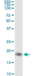
- Main image
- Experimental details
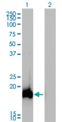
- Western Blot analysis of AHNAK expression in transfected 293T cell line by AHNAK monoclonal antibody (M01), clone 3G7.Lane 1: AHNAK transfected lysate(16 KDa).Lane 2: Non-transfected lysate.
Supportive validation
- Submitted by
- Abnova Corporation (provider)
- Main image
- Experimental details
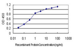
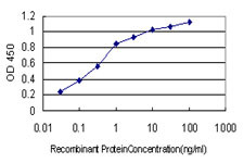
- Detection limit for recombinant GST tagged AHNAK is approximately 0.03ng/ml as a capture antibody.
- Validation comment
- Sandwich ELISA (Recombinant protein)
- Protocol
- Protocol
Supportive validation
- Submitted by
- Abnova Corporation (provider)
- Main image
- Experimental details
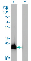
- Immunoprecipitation of AHNAK transfected lysate using anti-AHNAK monoclonal antibody and Protein A Magnetic Bead (U0007), and immunoblotted with AHNAK MaxPab rabbit polyclonal antibody.
- Validation comment
- Immunoprecipitation
- Protocol
- Protocol
