Explore
 Validate
 Learn
Learn Western blot
 Immunocytochemistry
Antibody data
- Antibody Data
- Antigen structure
- References [1]
- Comments [0]
- Validations
- Immunocytochemistry [4]
- Immunohistochemistry [1]
- Other assay [1]
Submit
Validation data
Reference
Comment
Report error
- Product number
- PA5-53890 - Provider product page

- Provider
- Invitrogen Antibodies
- Product name
- AHNAK Polyclonal Antibody
- Antibody type
- Polyclonal
- Antigen
- Recombinant protein fragment
- Description
- Immunogen sequence: LGEGHLSVKG SGGEWKGPQV SSALNLDTSK FAGGLHFSGP KVEGGVKGGQ IGLQAPGLSV SGPQGHLESG SGKVTFPKMK IPKFTFSGRE LVGREMGVDV HFPKAEASIQ AGAGDGEWEE SEVKLKKSKI KMPK Highest antigen sequence identity to the following orthologs: Mouse - 87%, Rat - 84%.
- Reactivity
- Human
- Host
- Rabbit
- Isotype
- IgG
- Vial size
- 100 μL
- Concentration
- 0.1 mg/mL
- Storage
- Store at 4°C short term. For long term storage, store at -20°C, avoiding freeze/thaw cycles.
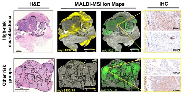
Submitted references Discovery of Spatial Peptide Signatures for Neuroblastoma Risk Assessment by MALDI Mass Spectrometry Imaging.
Wu Z, Hundsdoerfer P, Schulte JH, Astrahantseff K, Boral S, Schmelz K, Eggert A, Klein O
Cancers 2021 Jun 25;13(13)
Cancers 2021 Jun 25;13(13)
No comments: Submit comment
Supportive validation
- Submitted by
- Invitrogen Antibodies (provider)
- Main image
- Experimental details
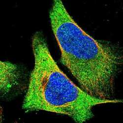
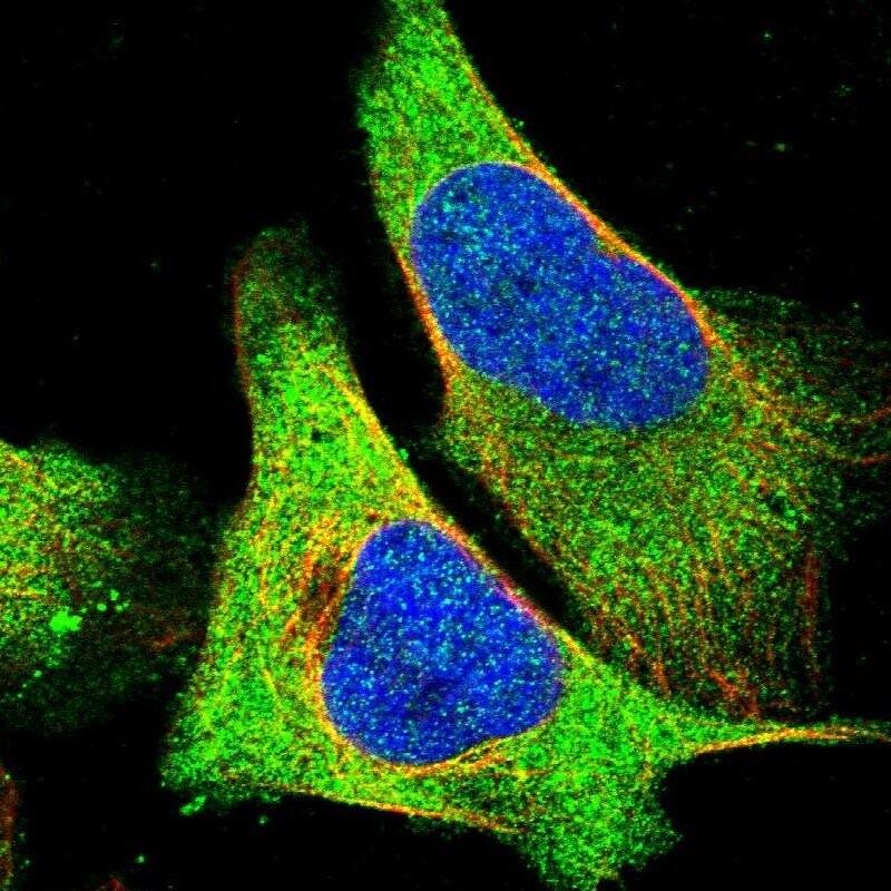
- Immunofluorescent staining of AHNAK in human cell line U-2 OS shows positivity in plasma membrane & cytoplasm. Samples were probed using an AHNAK Polyclonal Antibody (Product # PA5-53890).

- Submitted by
- Invitrogen Antibodies (provider)
- Main image

- Experimental details
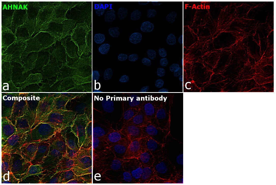
- Immunofluorescence analysis of AHNAK was performed using 70% confluent log phase HaCaT cells. The cells were fixed with 4% paraformaldehyde for 10 minutes, permeabilized with 0.1% Triton™ X-100 for 15 minutes, and blocked with 2% BSA for 1 hour at room temperature. The cells were labeled with AHNAK Polyclonal Antibody (Product # PA5-53890, 0.25 µg/mL) in 0.1% BSA, incubated at 4 degree celsius overnight and then labeled with Donkey anti-Rabbit IgG (H+L) Highly Cross-Adsorbed Secondary Antibody, Alexa Fluor™ Plus 488 (Product # A32790, 1:2000) for 45 minutes at room temperature (Panel a: Green). Nuclei (Panel b:Blue) were stained with ProLong™ Diamond Antifade Mountant with DAPI (Product # P36962). F-actin (Panel c: Red) was stained with Rhodamine Phalloidin (Product # R415, 1:300). Panel d represents the merged image showing cell membrane and cytosolic localization. Panel e represents control cells with no primary antibody to assess background. The images were captured at 60X magnification.
- Submitted by
- Invitrogen Antibodies (provider)
- Main image

- Experimental details
- Immunofluorecent analysis of AHNAK in human cell line U-2 OS using AHNAK Polyclonal Antibody (Product # PA5-53890). Staining shows localization to plasma membrane and cytosol.
- Submitted by
- Invitrogen Antibodies (provider)
- Main image

- Experimental details
- Immunofluorescence analysis of AHNAK was performed using 70% confluent log phase HaCaT cells. The cells were fixed with 4% paraformaldehyde for 10 minutes, permeabilized with 0.1% Triton™ X-100 for 15 minutes, and blocked with 2% BSA for 1 hour at room temperature. The cells were labeled with AHNAK Polyclonal Antibody (Product # PA5-53890, 0.25 µg/mL) in 0.1% BSA, incubated at 4 degree celsius overnight and then labeled with Donkey anti-Rabbit IgG (H+L) Highly Cross-Adsorbed Secondary Antibody, Alexa Fluor™ Plus 488 (Product # A32790, 1:2000) for 45 minutes at room temperature (Panel a: Green). Nuclei (Panel b:Blue) were stained with ProLong™ Diamond Antifade Mountant with DAPI (Product # P36962). F-actin (Panel c: Red) was stained with Rhodamine Phalloidin (Product # R415, 1:300). Panel d represents the merged image showing cell membrane and cytosolic localization. Panel e represents control cells with no primary antibody to assess background. The images were captured at 60X magnification.

Supportive validation
- Submitted by
- Invitrogen Antibodies (provider)
- Main image

- Experimental details
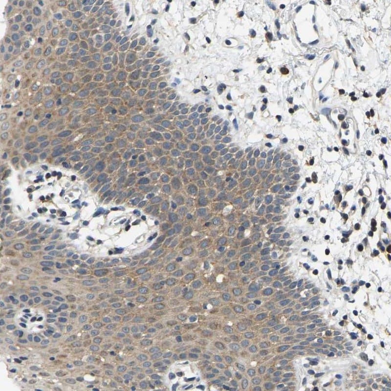
- Immunohistochemical analysis of AHNAK in human oral mucosa using AHNAK Polyclonal Antibody (Product # PA5-53890) shows cytoplasmic positivity in squamous epithelial cells.
Supportive validation
- Submitted by
- Invitrogen Antibodies (provider)
- Main image
- Experimental details
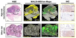
- Figure 3 Validation of two discriminative protein markers for neuroblastoma risk in tissue sections. Shown are representative tissue sections from neuroblastoma designated high-risk (HR) and as other risk groups (nHR). MALDI-MSI ion maps for one peptide (m/z 1832.79 Da) assigned to AHNAK and one peptide (m/z 922.50 Da) assigned to CRMP1 are shown next to the corresponding sections stained with hematoxylin and eosin (H&E) for orientation. Black lines border areas with >80% tumor cell content. Immunohistochemical (IHC) detection of AHNAK and CRMP1 is shown for the regions surrounded by the yellow squares in the expanded image (400x magnification).