Explore
 Validate
 Learn
Learn Western blot
 Immunocytochemistry
 Immunoprecipitation
Antibody data
- Antibody Data
- Antigen structure
- References [1]
- Comments [0]
- Validations
- Immunocytochemistry [8]
- Immunohistochemistry [1]
- Other assay [2]
Submit
Validation data
Reference
Comment
Report error
- Product number
- MA1-10050 - Provider product page

- Provider
- Invitrogen Antibodies
- Product name
- AHNAK Monoclonal Antibody (EM-09)
- Antibody type
- Monoclonal
- Antigen
- Other
- Description
- This antibody reacts with AHNAK1, a 700 kDa multi-functional adaptor protein expressed mainly in epithelial cell, various types of muscle cells and immune cells. Immunocytochemistry: permeabilization is required.
- Reactivity
- Human, Mouse
- Host
- Mouse
- Isotype
- IgG
- Antibody clone number
- EM-09
- Vial size
- 100 μg
- Concentration
- 1 mg/mL
- Storage
- 4°C, do not freeze
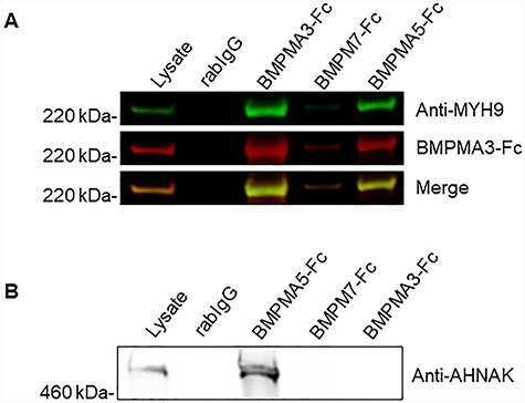
Submitted references A yeast display immunoprecipitation screen for targeted discovery of antibodies against membrane protein complexes.
Lajoie JM, Cho YK, Frost D, Bremner S, Li L, Shusta EV
Protein engineering, design & selection : PEDS 2019 Dec 31;32(5):219-230
Protein engineering, design & selection : PEDS 2019 Dec 31;32(5):219-230
No comments: Submit comment
Supportive validation
- Submitted by
- Invitrogen Antibodies (provider)
- Main image

- Experimental details
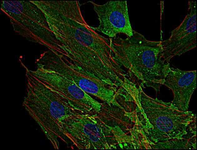
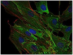
- Immunofluorescence staining of AHNAK1 in human primary fibroblasts using anti-AHNAK1 (EM-09; green). Actin filaments were decorated by phalloidin (red) and cell nuclei stained with DAPI (blue).
- Submitted by
- Invitrogen Antibodies (provider)
- Main image
- Experimental details
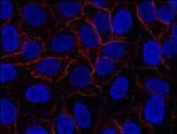
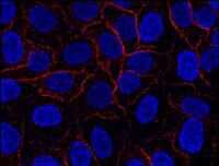
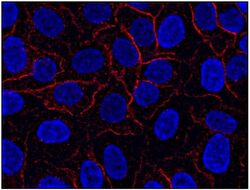
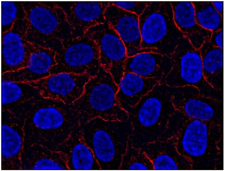
- Immunofluorescence staining of AHNAK1 in HeLa cell line using anti-AHNAK1 (EM-09; red). Cell nuclei stained with DAPI (blue).
- Submitted by
- Invitrogen Antibodies (provider)
- Main image
- Experimental details
- Immunocytochemistry staining of AHNAK1 in HeLa cell line using anti-AHNAK1 (EM-09; red) Monoclonal antibody (Product # MA1-10050). Cell nuclei stained with DAPI (blue).
- Submitted by
- Invitrogen Antibodies (provider)
- Main image
- Experimental details
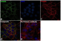
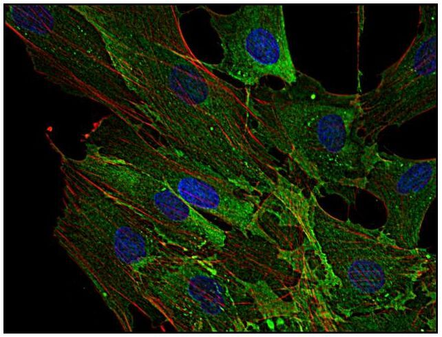
- Immunofluorescence analysis of AHNAK was performed using 70% confluent log phase HaCaT cells. The cells were fixed with 4% paraformaldehyde for 10 minutes, permeabilized with 0.1% Triton™ X-100 for 15 minutes, and blocked with 2% BSA for 1 hour at room temperature. The cells were labeled with AHNAK Monoclonal Antibody (EM-09) (Product # MA1-10050, 1:100) in 0.1% BSA, incubated at 4 degree celsius overnight and then labeled with Donkey anti-Mouse IgG (H+L) Highly Cross-Adsorbed Secondary Antibody, Alexa Fluor™ Plus 488 (Product # A32766, 1:2000), for 45 minutes at room temperature (Panel a: Green). Nuclei (Panel b:Blue) were stained with ProLong™ Diamond Antifade Mountant with DAPI (Product # P36962). F-actin (Panel c: Red) was stained with Rhodamine Phalloidin (Product # R415, 1:300). Panel d represents the merged image showing cell membrane and cytosolic localization. Panel e represents control cells with no primary antibody to assess background. The images were captured at 60X magnification.

- Submitted by
- Invitrogen Antibodies (provider)
- Main image
- Experimental details
- Immunocytochemistry staining of AHNAK1 in primary human fibroblasts using anti-AHNAK1 (EM-09; green) Monoclonal antibody (Product # MA1-10050). Actin filaments were decorated by phalloidin (red) and cell nuclei stained with DAPI (blue).
- Submitted by
- Invitrogen Antibodies (provider)
- Main image

- Experimental details
- Immunocytochemistry staining of AHNAK1 in HeLa cell line using anti-AHNAK1 (EM-09; red) Monoclonal antibody (Product # MA1-10050). Cell nuclei stained with DAPI (blue).

- Submitted by
- Invitrogen Antibodies (provider)
- Main image

- Experimental details
- Immunocytochemistry staining of AHNAK1 in primary human fibroblasts using anti-AHNAK1 (EM-09; green) Monoclonal antibody (Product # MA1-10050). Actin filaments were decorated by phalloidin (red) and cell nuclei stained with DAPI (blue).

- Submitted by
- Invitrogen Antibodies (provider)
- Main image

- Experimental details
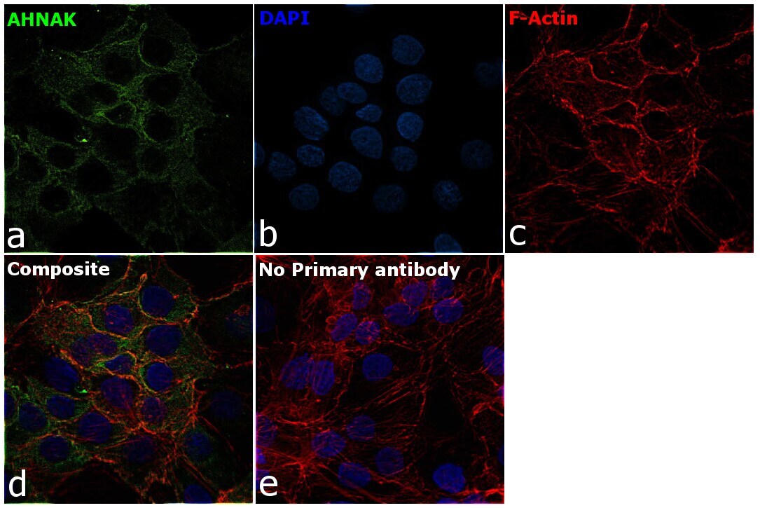
- Immunofluorescence analysis of AHNAK was performed using 70% confluent log phase HaCaT cells. The cells were fixed with 4% paraformaldehyde for 10 minutes, permeabilized with 0.1% Triton™ X-100 for 15 minutes, and blocked with 2% BSA for 1 hour at room temperature. The cells were labeled with AHNAK Monoclonal Antibody (EM-09) (Product # MA1-10050, 1:100) in 0.1% BSA, incubated at 4 degree celsius overnight and then labeled with Donkey anti-Mouse IgG (H+L) Highly Cross-Adsorbed Secondary Antibody, Alexa Fluor™ Plus 488 (Product # A32766, 1:2000), for 45 minutes at room temperature (Panel a: Green). Nuclei (Panel b:Blue) were stained with ProLong™ Diamond Antifade Mountant with DAPI (Product # P36962). F-actin (Panel c: Red) was stained with Rhodamine Phalloidin (Product # R415, 1:300). Panel d represents the merged image showing cell membrane and cytosolic localization. Panel e represents control cells with no primary antibody to assess background. The images were captured at 60X magnification.
Supportive validation
- Submitted by
- Invitrogen Antibodies (provider)
- Main image
- Experimental details
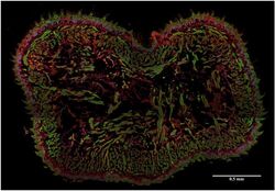
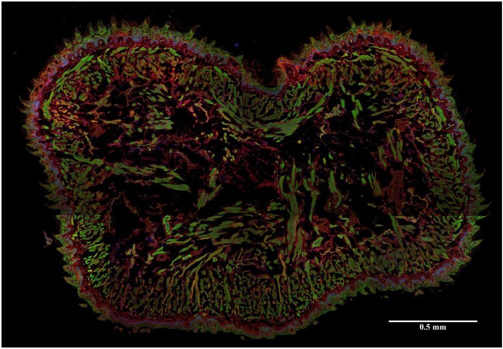
- Immunohistochemistry (Frozen) analysis of AHNAK using AHNAK Monoclonal Antibody (EM-09) (Product # MA1-10050) in murine tongue (EM-09; red). Actin filaments were decorated by phalloidin (green), cell nuclei stained with DAPI (blue).
Supportive validation
- Submitted by
- Invitrogen Antibodies (provider)
- Main image

- Experimental details
- NULL
- Submitted by
- Invitrogen Antibodies (provider)
- Main image

- Experimental details
- NULL