Explore
 Validate
 Learn
Learn Western blot
Antibody data
- Antibody Data
- Antigen structure
- References [0]
- Comments [0]
- Validations
- Western blot [5]
- Immunocytochemistry [3]
- Immunohistochemistry [1]
Submit
Validation data
Reference
Comment
Report error
- Product number
- PA5-78171 - Provider product page

- Provider
- Invitrogen Antibodies
- Product name
- Fascin Polyclonal Antibody
- Antibody type
- Polyclonal
- Antigen
- Recombinant full-length protein
- Description
- Positive Control: HeLa Predicted Reactivity: Rat (97%), Pig (94%), Bovine (94%) Store product as a concentrated solution. Centrifuge briefly prior to opening the vial.
- Reactivity
- Human, Mouse, Rat
- Host
- Rabbit
- Isotype
- IgG
- Vial size
- 100 µL
- Concentration
- 1.0 mg/mL
- Storage
- Store at 4°C short term. For long term storage, store at -20°C, avoiding freeze/thaw cycles.
No comments: Submit comment
Supportive validation
- Submitted by
- Invitrogen Antibodies (provider)
- Main image
- Experimental details
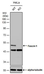
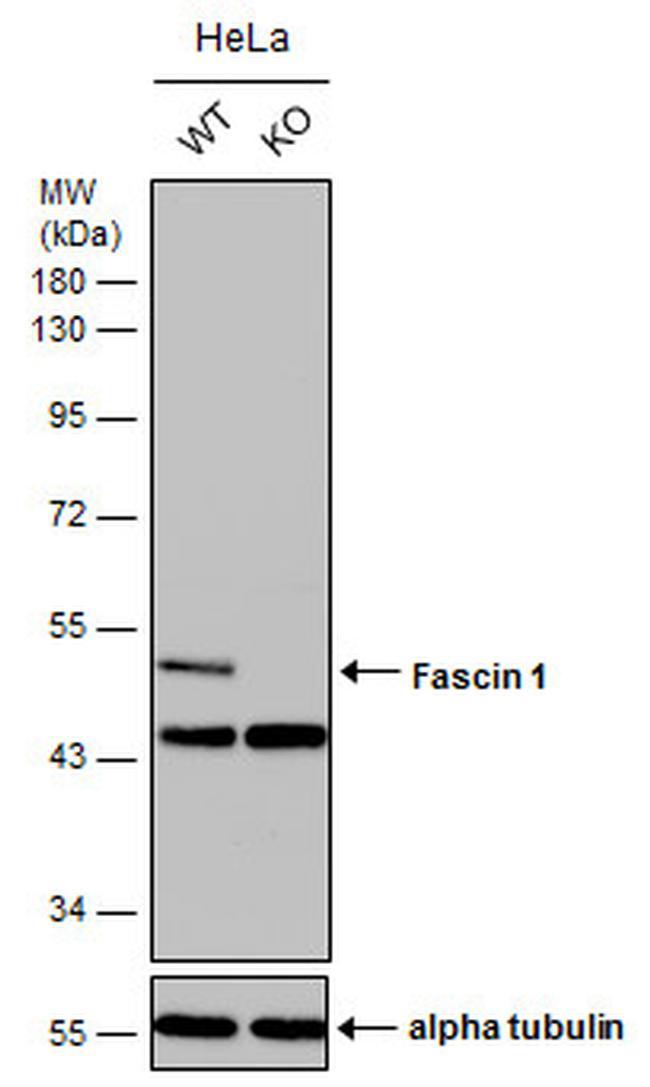
- Western blot analysis of Fascin 1 in wild-type and Fascin 1 knockout HeLa cells using 30 µg of protein. Samples were separated with 10% SDS-PAGE and incubated with Fascin 1 polyclonal antibody (Product # PA5-78171) using a dilution of 1:2000 followed by HRP-conjugated anti-rabbit IgG.
- Submitted by
- Invitrogen Antibodies (provider)
- Main image
- Experimental details
- Western blot analysis of Fascin 1 in HeLa cells using 30 µg of protein. Samples were separated with 10% SDS-PAGE and incubated with Fascin 1 polyclonal antibody (Product # PA5-78171) using a dilution of 1:1000.

- Submitted by
- Invitrogen Antibodies (provider)
- Main image

- Experimental details
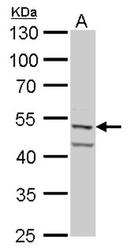
- Fascin Polyclonal Antibody detects Fascin 1 protein by Western blot analysis. A. 30 µg HeLa whole cell lysate/extract.10 % SDS-PAGE. Fascin Polyclonal Antibody (Product # PA5-78171) dilution: 1:1,000.

- Submitted by
- Invitrogen Antibodies (provider)
- Main image
- Experimental details
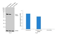
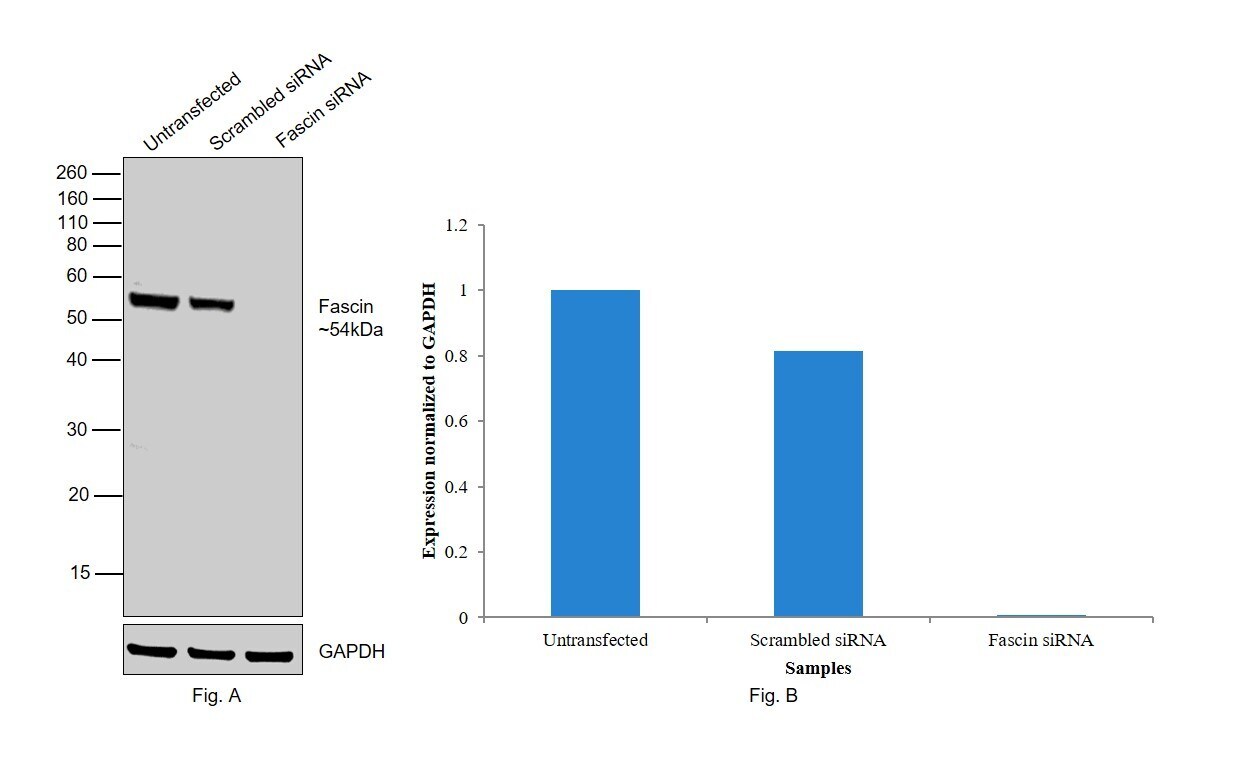
- Knockdown of Fascin was achieved by transfecting SH-SY5Y with Fascin specific siRNAs (Silencer® select Product # s13208, s13207). Western blot analysis (Fig. a) was performed using Whole cell extracts from the Fascin knockdown cells (lane 3), non-targeting scrambled siRNA transfected cells (lane 2) and untransfected cells (lane 1). The blot was probed with Fascin Polyclonal Antibody (Product # PA5-78171, 1:1000 ) and Goat anti-Rabbit IgG (H+L) Superclonal™ Recombinant Secondary Antibody, HRP (Product # A27036, 1:4000). Densitometric analysis of this western blot is shown in histogram (Fig. b). Loss of signal upon siRNA mediated knock down confirms that antibody is specific to Fascin.
- Submitted by
- Invitrogen Antibodies (provider)
- Main image

- Experimental details
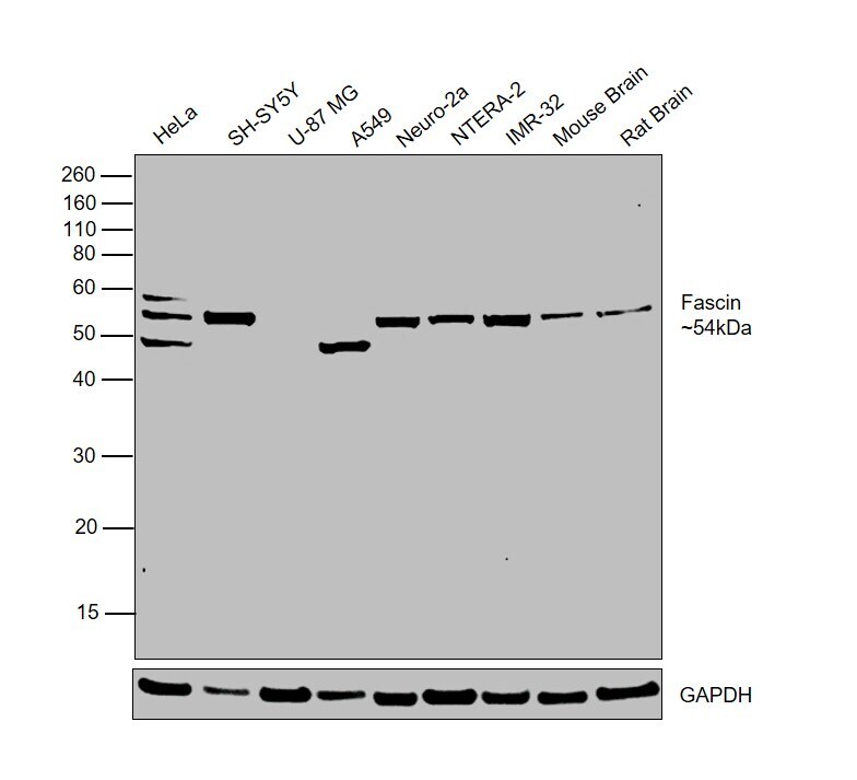
- Western blot was performed using Anti-Fascin Polyclonal Antibody(Product # PA5-78171) and a 54kDa band corresponding to Fascin was observed across all the tested cell lines and tissues, except U-87 MG and A549. Whole cell extracts (30 µg lysate) of HeLa (Lane 1), SH-SY5Y (Lane 2), U-87 MG (Lane 3), A549 (Lane 4), Neuro-2a (Lane 5), NTERA-2 cl.D1 (Lane 6), IMR-32 (Lane 7), Mouse Brain (Lane 8), Rat Brain (Lane 9) were electrophoresed using NuPAGE™ 10% Bis-Tris Protein Gel (Product # NP0302BOX). Resolved proteins were then transferred onto a Nitrocellulose membrane (Product # IB23001) by iBlot® 2 Dry Blotting System (Product # IB21001). The blot was probed with the primary antibody (1:1000) and detected by chemiluminescence with Goat anti-Rabbit IgG (H+L) Superclonal™ Recombinant Secondary Antibody, HRP (Product # A27036, 1:4000) using the iBright FL 1000 (Product # A32752). Chemiluminescent detection was performed using Novex® ECL Chemiluminescent Substrate Reagent Kit (Product # WP20005).
Supportive validation
- Submitted by
- Invitrogen Antibodies (provider)
- Main image
- Experimental details
- Immunofluorescent analysis of Fascin 1 in HCT116 cells. Samples were treated with 100% MeOH for 5 min and incubated with Fascin 1 polyclonal antibody (Product # PA5-78171) using a dilution of 1:500, followed by Hoechst.
- Submitted by
- Invitrogen Antibodies (provider)
- Main image
- Experimental details
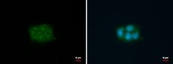
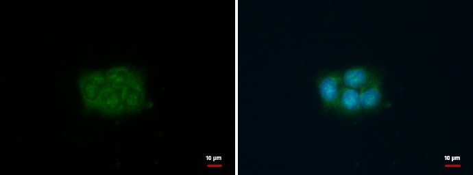
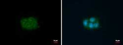
- FSCN1 antibody detects FSCN1 protein at cytoplasm by immunofluorescent analysis. Sample: HCT116 cells were fixed in 100% MeOH for 5 min. Green: FSCN1 protein stained by FSCN1 antibody (Product # PA5-78171) diluted at 1:500. Blue: Hoechst 33342 staining.
- Submitted by
- Invitrogen Antibodies (provider)
- Main image
- Experimental details
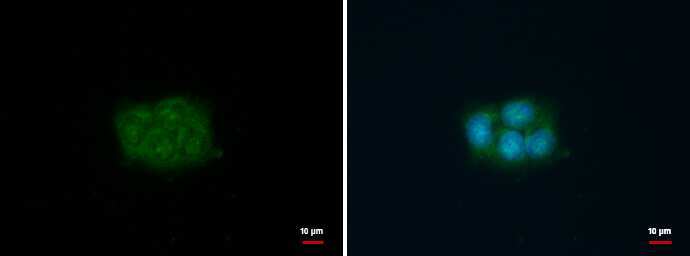
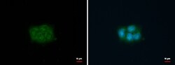
- FSCN1 antibody detects FSCN1 protein at cytoplasm by immunofluorescent analysis. Sample: HCT116 cells were fixed in 100% MeOH for 5 min. Green: FSCN1 protein stained by FSCN1 antibody (Product # PA5-78171) diluted at 1:500. Blue: Hoechst 33342 staining.

Supportive validation
- Submitted by
- Invitrogen Antibodies (provider)
- Main image

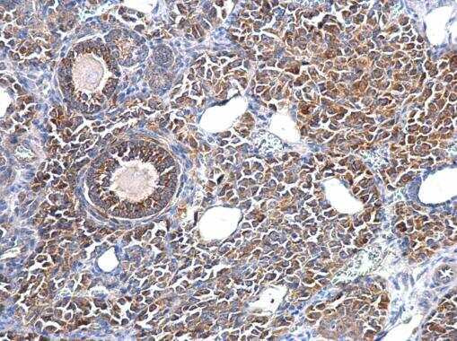
- Experimental details
- Fascin Polyclonal Antibody detects Fascin 1 protein at cytosol on mouse ovary by immunohistochemical analysis. Sample: Paraffin-embedded mouse ovary. Fascin Polyclonal Antibody (Product # PA5-78171) dilution: 1:500. Antigen Retrieval: EDTA based buffer, pH 8.0, 15 min.