 Explore
 Validate
 Learn
Learn Western blot
Antibody data
- Antibody Data
- Antigen structure
- References [9]
- Comments [0]
- Validations
- Western blot [2]
- Immunohistochemistry [1]
- Other assay [3]
Submit
Validation data
Reference
Comment
Report error
- Product number
- MA5-11483 - Provider product page

- Provider
- Invitrogen Antibodies
- Product name
- Fascin Monoclonal Antibody (FCN01 (55K-2))
- Antibody type
- Monoclonal
- Antigen
- Purifed from natural sources
- Description
- eFluor® 450 is an alternative to Pacific Blue®. eFluor® 450 emits at 445 nm and is excited with the Violet laser (405 nm). Please make sure that your instrument is capable of detecting this fluorochome.
- Reactivity
- Human, Mouse, Rat
- Host
- Mouse
- Isotype
- IgG
- Antibody clone number
- FCN01 (55K-2)
- Vial size
- 500 µL
- Concentration
- Conc. Not Determined
- Storage
- 4° C
Submitted references Association of Fascin and matrix metalloproteinase-9 expression with poor prognostic parameters in breast carcinoma of Egyptian women.
Fascin expression predicts lymph node metastasis and worse survival in small intestinal carcinoma.
The correlation of cortactin and fascin-1 expression with clinicopathological parameters in pancreatic and ampulla of Vater adenocarcinoma.
EGFR signaling regulates tumor cell migration in craniopharyngiomas.
Fascin expression and its potential significance in gastrointestinal stromal tumors.
Fascin expression and its potential significance in gastrointestinal stromal tumors.
Prognostic significance of fascin-1 and E-cadherin expression in laryngeal squamous cell carcinoma.
Availability of activated CD4+ T cells dictates the level of viremia in naturally SIV-infected sooty mangabeys.
Proteomic identification of novel proteins in cortical lewy bodies.
Youssef NS, Hakim SA
Diagnostic pathology 2014 Jul 4;9:136
Diagnostic pathology 2014 Jul 4;9:136
Fascin expression predicts lymph node metastasis and worse survival in small intestinal carcinoma.
Gu MJ, Kim JY, Park JB
Pathology 2014 Jan;46(1):21-4
Pathology 2014 Jan;46(1):21-4
The correlation of cortactin and fascin-1 expression with clinicopathological parameters in pancreatic and ampulla of Vater adenocarcinoma.
Tsai WC, Lin CK, Lee HS, Gao HW, Nieh S, Chan DC, Jin JS
APMIS : acta pathologica, microbiologica, et immunologica Scandinavica 2013 Mar;121(3):171-81
APMIS : acta pathologica, microbiologica, et immunologica Scandinavica 2013 Mar;121(3):171-81
EGFR signaling regulates tumor cell migration in craniopharyngiomas.
Hölsken A, Gebhardt M, Buchfelder M, Fahlbusch R, Blümcke I, Buslei R
Clinical cancer research : an official journal of the American Association for Cancer Research 2011 Jul 1;17(13):4367-77
Clinical cancer research : an official journal of the American Association for Cancer Research 2011 Jul 1;17(13):4367-77
Fascin expression and its potential significance in gastrointestinal stromal tumors.
Ozcan A, Karslioğlu Y, Günal A, Cermık AH, Kurt B, Ongürü O
The Turkish journal of gastroenterology : the official journal of Turkish Society of Gastroenterology 2011 Aug;22(4):363-8
The Turkish journal of gastroenterology : the official journal of Turkish Society of Gastroenterology 2011 Aug;22(4):363-8
Prognostic significance of fascin-1 and E-cadherin expression in laryngeal squamous cell carcinoma.
Zou J, Yang H, Chen F, Zhao H, Lin P, Zhang J, Ye H, Wang L, Liu S
European journal of cancer prevention : the official journal of the European Cancer Prevention Organisation (ECP) 2010 Jan;19(1):11-7
European journal of cancer prevention : the official journal of the European Cancer Prevention Organisation (ECP) 2010 Jan;19(1):11-7
Availability of activated CD4+ T cells dictates the level of viremia in naturally SIV-infected sooty mangabeys.
Klatt NR, Villinger F, Bostik P, Gordon SN, Pereira L, Engram JC, Mayne A, Dunham RM, Lawson B, Ratcliffe SJ, Sodora DL, Else J, Reimann K, Staprans SI, Haase AT, Estes JD, Silvestri G, Ansari AA
The Journal of clinical investigation 2008 Jun;118(6):2039-49
The Journal of clinical investigation 2008 Jun;118(6):2039-49
Proteomic identification of novel proteins in cortical lewy bodies.
Leverenz JB, Umar I, Wang Q, Montine TJ, McMillan PJ, Tsuang DW, Jin J, Pan C, Shin J, Zhu D, Zhang J
Brain pathology (Zurich, Switzerland) 2007 Apr;17(2):139-45
Brain pathology (Zurich, Switzerland) 2007 Apr;17(2):139-45
No comments: Submit comment
Supportive validation
- Submitted by
- Invitrogen Antibodies (provider)
- Main image

- Experimental details
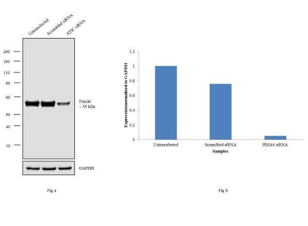
- Knockdown of Fascin was achieved by transfecting SHSY-5Y cells with Fascin specific siRNAs (Silencer® select Product # s13207, s13208). Western blot analysis (Fig a) was performed using whole cell extracts from the Fascin knock down cells (lane 3), non-specific scrambled siRNA transfected cells (lane 2) and untransfected cells (lane 1). The blots were probed with anti-Fascin Mouse monoclonal Antibody (Product # MA5-11483, 1:500 dilution) and Goat anti-Mouse IgG (H+L) Superclonal™ Secondary Antibody, HRP conjugate (Product # A28177, 0.25 µg/mL, 1:4000 dilution). Densitometric analysis of this western blot is shown in histogram (Fig b). Decrease in signal upon siRNA mediated knock down confirms that antibody is specific to Fascin.
- Submitted by
- Invitrogen Antibodies (provider)
- Main image

- Experimental details
- Western blot analysis was performed on whole cell extracts (30 µg lysate) of HeLa (Lane 1), SH-SY5Y (Lane 2), U-87 MG (Lane 3), SK-N-AS (Lane 4), A549 (Lane 5), A-431 (Lane 6), Neuro-2a (Lane 7), NTERA-2 (Lane 8), IMR-32 (Lane 9) and tissue extract of Rat Brain (Lane 10). The blots were probed with Anti-Fascin Mouse Monoclonal Antibody (Product # MA5-11483, 1:250 dilution) and detected by chemiluminescence using Goat anti-Mouse IgG (H+L) Superclonal™ Secondary Antibody, HRP conjugate (Product # A28177, 0.4 µg/mL, 1:2500 dilution). A 55 kDa band corresponding to Fascin was observed across the cell lines and tissue tested. Known quantity of protein samples were electrophoresed using Novex® NuPAGE® 4-12 % Bis-Tris gel (Product # NP0321BOX), XCell SureLock™ Electrophoresis System (Product # EI0002) and Novex® Sharp Pre-Stained Protein Standard (Product # LC5800). Resolved proteins were then transferred onto a nitrocellulose membrane with iBlot® 2 Dry Blotting System (Product # IB21001). The membrane was probed with the relevant primary and secondary Antibody following blocking with 5 % skimmed milk. Chemiluminescent detection was performed using Pierce™ ECL Western Blotting Substrate (Product # 32106).

Supportive validation
- Submitted by
- Invitrogen Antibodies (provider)
- Main image
- Experimental details
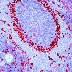
- Formalin-fixed, paraffin-embedded rat spleen stained with Fascin antibody using peroxidase-conjugate and AEC chromogen. Note cytoplasmic staining of splenic cells.

Supportive validation
- Submitted by
- Invitrogen Antibodies (provider)
- Main image
- Experimental details
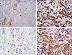
- Figure 1 Fascin and MMP-9 expressions in normal breast tissue and invasive ductal carcinomas. a : Moderate fascin expression in the myoepithelial cells and luminal cells of few normal acini (IHC x 400). b : Positive cytoplasmic fascin expression in malignant cells (IHC x 400). c : Negative MMP-9 expression in normal breast tissue (IHC x 400). d : Positive cytoplasmic MMP-9 expression of malignant cells with weak expression of stromal cells (IHC x 400).

- Submitted by
- Invitrogen Antibodies (provider)
- Main image

- Experimental details
- Figure 2 Fascin and MMP-9 expression in invasive ductal carcinomas with adjacent in situ component. (a) : Positive cytoplasmic fascin expression in the myoepithelial cells of the in situ component (IHC x 200). (b) : Positive cytoplasmic fascin expression in the in situ carcinoma lesions as well as the adjacent invasive carcinoma (IHC x 200). (c) : Positive cytoplasmic MMP-9 expression in the in situ carcinoma lesions (IHC x 200). (d) : Positive cytoplasmic MMP-9 expression in invasive carcinoma and negative expression in the in situ carcinoma lesions (IHC x 200).

- Submitted by
- Invitrogen Antibodies (provider)
- Main image

- Experimental details
- Figure 5 Invasive ductal breast carcinomas. a : Enhanced fascin expression at the leading edge of the tumor (IHC x 100). b : The same field with higher magnification power showing positive cytoplasmic fascin expression of malignant cells and endothelial cells (IHC x 200). c : Enhanced MMP-9 expression at the tumor-host border (IHC x 100). d : The same field with higher magnification power showing positive cytoplasmic MMP-9 expression of malignant cells (IHC x 200).
