Explore
 Validate
 Learn
Learn Western blot
Antibody data
- Antibody Data
- Antigen structure
- References [0]
- Comments [0]
- Validations
- Western blot [2]
Submit
Validation data
Reference
Comment
Report error
- Product number
- AF7745 - Provider product page

- Provider
- R&D Systems
- Product name
- Human/Mouse/Rat Fascin Antibody
- Antibody type
- Polyclonal
- Description
- Antigen Affinity-purified. Detects human, mouse, rat Fascin in direct ELISAs and Western blots. In direct ELISAs, less than 5% cross-reactivity with recombinant human Fascin-2 is observed.
- Reactivity
- Human, Mouse, Rat
- Host
- Sheep
- Conjugate
- Unconjugated
- Antigen sequence
Q16658- Isotype
- IgG
- Vial size
- 100 ug
- Concentration
- LYOPH
- Storage
- Use a manual defrost freezer and avoid repeated freeze-thaw cycles. 12 months from date of receipt, -20 to -70 °C as supplied. 1 month, 2 to 8 °C under sterile conditions after reconstitution. 6 months, -20 to -70 °C under sterile conditions after reconstitution.
No comments: Submit comment
Supportive validation
- Submitted by
- R&D Systems (provider)
- Main image
- Experimental details
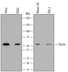
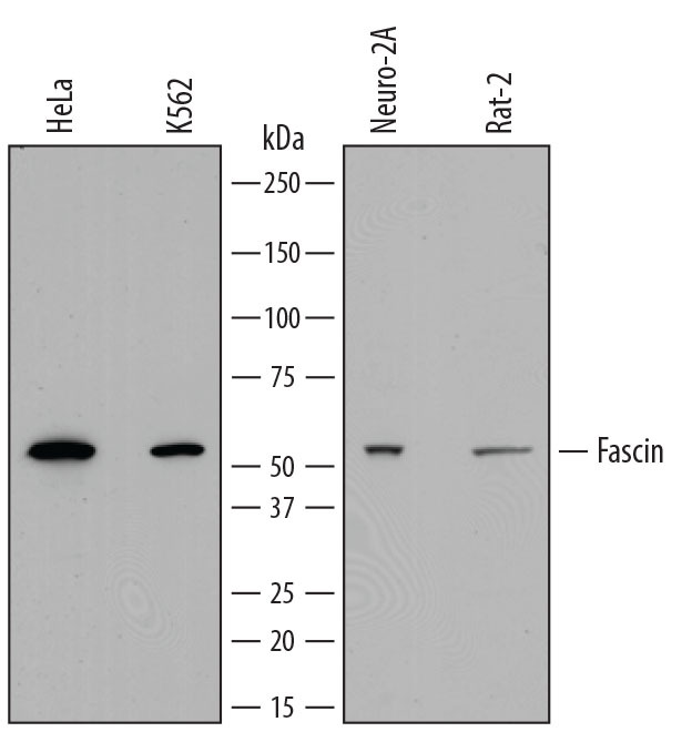
- Detection of Human, Mouse, and Rat Fascin by Western Blot. Western blot shows lysates of HeLa human cervical epithelial carcinoma cell line, K562 human chronic myelogenous leukemia cell line, Neuro-2A mouse neuroblastoma cell line, and Rat-2 rat thymidine kinase-deficient embryonic fibroblast cell line. PVDF membrane was probed with 0.4 µg/mL of Sheep Anti-Human/Mouse/Rat Fascin Antigen Affinity-purified Polyclonal Antibody (Catalog # AF7745) followed by HRP-conjugated Anti-Sheep IgG Secondary Antibody (Catalog # HAF016). A specific band was detected for Fascin at approximately 55 kDa (as indicated). This experiment was conducted under reducing conditions using Immunoblot Buffer Group 1.
- Submitted by
- R&D Systems (provider)
- Main image
- Experimental details
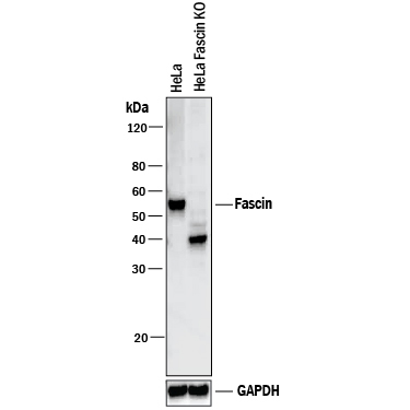
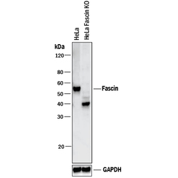
- Western Blot Shows Human Fascin Specificity by Using Knockout Cell Line. Western blot shows lysates of HeLa human cervical epithelial carcinoma parental cell line and Fascin knockout HeLa cell line (KO). PVDF membrane was probed with 0.4 µg/mL of Sheep Anti-Human/Mouse/Rat Fascin Antigen Affinity-purified Polyclonal Antibody (Catalog # AF7745) followed by HRP-conjugated Anti-Sheep IgG Secondary Antibody (Catalog # HAF016). A specific band was detected for Fascin at approximately 55 kDa (as indicated) in the parental HeLa cell line, but is not detectable in knockout HeLa cell line. GAPDH (Catalog # AF5718) is shown as a loading control. This experiment was conducted under reducing conditions and using Immunoblot Buffer Group 1. New adjunct appears with knockout cell line.