Explore
 Validate
 Learn
Learn Western blot
Antibody data
- Antibody Data
- Antigen structure
- References [0]
- Comments [0]
- Validations
- Western blot [3]
- Immunohistochemistry [1]
Submit
Validation data
Reference
Comment
Report error
- Product number
- MAB7745 - Provider product page

- Provider
- R&D Systems
- Product name
- Human Fascin Antibody
- Antibody type
- Monoclonal
- Description
- Protein A or G purified from hybridoma culture supernatant. Detects human Fascin in ELISAs and Western blots. Detects mouse and rat Fascin in Western blots. In direct ELISAs, no cross-reactivity with recombinant human Fascin-2 is observed.
- Reactivity
- Human
- Host
- Mouse
- Conjugate
- Unconjugated
- Antigen sequence
Q16658- Isotype
- IgG
- Antibody clone number
- 833223
- Vial size
- 100 ug
- Concentration
- LYOPH
- Storage
- Use a manual defrost freezer and avoid repeated freeze-thaw cycles. 12 months from date of receipt, -20 to -70 °C as supplied. 1 month, 2 to 8 °C under sterile conditions after reconstitution. 6 months, -20 to -70 °C under sterile conditions after reconstitution.
No comments: Submit comment
Supportive validation
- Submitted by
- R&D Systems (provider)
- Main image
- Experimental details
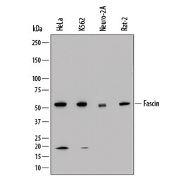
- Detection of Human, Mouse, and Rat Fascin by Western Blot. Western blot shows lysates of HeLa human cervical epithelial carcinoma cell line, K562 human chronic myelogenous leukemia cell line, Neuro-2A mouse neuroblastoma cell line, and Rat-2 rat embryonic fibroblast cell line. PVDF membrane was probed with 1 µg/mL of Mouse Anti-Human Fascin Monoclonal Antibody (Catalog # MAB7745) followed by HRP-conjugated Anti-Mouse IgG Secondary Antibody (Catalog # HAF018). A specific band was detected for Fascin at approximately 55 kDa (as indicated). This experiment was conducted under reducing conditions and using Immunoblot Buffer Group 1.

- Submitted by
- R&D Systems (provider)
- Main image
- Experimental details
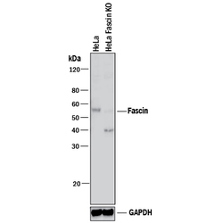
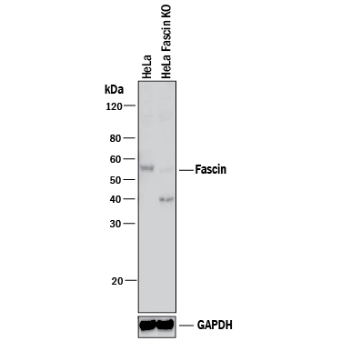
- Western Blot Shows Human Fascin Specificity by Using Knockout Cell Line. Western blot shows lysates of HeLa human cervical epithelial carcinoma parental cell line and Fascin knockout HeLa cell line (KO). PVDF membrane was probed with 1 µg/mL of Mouse Anti-Human Fascin Monoclonal Antibody (Catalog # MAB7745) followed by HRP-conjugated Anti-Mouse IgG Secondary Antibody (Catalog # HAF018). A specific band was detected for Fascin at approximately 55 kDa (as indicated) in the parental HeLa cell line, but is not detectable in knockout HeLa cell line. GAPDH (Catalog # MAB5718) is shown as a loading control. This experiment was conducted under reducing conditions and using Immunoblot Buffer Group 1. New adjunct appears with knockout cell line.
- Submitted by
- R&D Systems (provider)
- Main image
- Experimental details
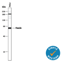
- Detection of Human Fascin by Simple WesternTM. Simple Western lane view shows lysates of HeLa human cervical epithelial carcinoma cell line, loaded at 0.5 mg/mL. A specific band was detected for Fascin at approximately 61 kDa (as indicated) using 10 µg/mL of Mouse Anti-Human Fascin Monoclonal Antibody (Catalog # MAB7745). This experiment was conducted under reducing conditions and using the 12-230 kDa separation system.

Supportive validation
- Submitted by
- R&D Systems (provider)
- Main image

- Experimental details
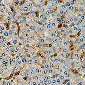
- Fascin in Human Liver. Fascin was detected in immersion fixed paraffin-embedded sections of human liver using Mouse Anti-Human Fascin Monoclonal Antibody (Catalog # MAB7745) at 15 µg/mL overnight at 4 °C. Before incubation with the primary antibody, tissue was subjected to heat-induced epitope retrieval using Antigen Retrieval Reagent-Basic (Catalog # CTS013). Tissue was stained using the Anti-Mouse HRP-DAB Cell & Tissue Staining Kit (brown; Catalog # CTS002) and counterstained with hematoxylin (blue). Specific staining was localized to synusoids. View our protocol for Chromogenic IHC Staining of Paraffin-embedded Tissue Sections.