 Explore
 Validate
 Learn
Learn Western blot
 Immunocytochemistry
Antibody data
- Antibody Data
- Antigen structure
- References [6]
- Comments [0]
- Validations
- Immunocytochemistry [1]
Submit
Validation data
Reference
Comment
Report error
- Product number
- HPA005723 - Provider product page

- Provider
- Atlas Antibodies
- Proper citation
- Atlas Antibodies Cat#HPA005723, RRID:AB_1849159
- Product name
- Anti-FSCN1
- Antibody type
- Polyclonal
- Description
- Polyclonal Antibody against Human FSCN1, Gene description: fascin actin-bundling protein 1, Alternative Gene Names: FLJ38511, p55, SNL, Validated applications: WB, IHC, ICC, Uniprot ID: Q16658, Storage: Store at +4°C for short term storage. Long time storage is recommended at -20°C.
- Reactivity
- Human, Mouse, Rat
- Host
- Rabbit
- Conjugate
- Unconjugated
- Isotype
- IgG
- Vial size
- 100 µl
- Concentration
- 0.1 mg/ml
- Storage
- Store at +4°C for short term storage. Long time storage is recommended at -20°C.
- Handling
- The antibody solution should be gently mixed before use.
Submitted references The combined tumour-based Fascin/Snail and stromal periostin reveals the effective prognosis prediction in colorectal cancer patients.
GRHL3 activates FSCN1 to relax cell-cell adhesions between migrating keratinocytes during wound reepithelialization
Survey of cancer cell anatomy in nonadhesive confinement reveals a role for filamin-A and fascin-1 in leader bleb–based migration
Sphingosine Kinase 1 Signaling Promotes Metastasis of Triple-Negative Breast Cancer
Single-cell transcriptomics reveals multi-step adaptations to endocrine therapy
Studying the role of fascin-1 in mechanically stressed podocytes
Jirapongwattana N, Thongchot S, Pongpaibul A, Trakarnsanga A, Quinn J, Thuwajit P, Thuwajit C, Edwards J
PloS one 2024;19(6):e0304666
PloS one 2024;19(6):e0304666
GRHL3 activates FSCN1 to relax cell-cell adhesions between migrating keratinocytes during wound reepithelialization
Kashgari G, Venkatesh S, Refuerzo S, Pham B, Bayat A, Klein R, Ramos R, Ta A, Plikus M, Wang P, Andersen B
JCI Insight 2021;6(17)
JCI Insight 2021;6(17)
Survey of cancer cell anatomy in nonadhesive confinement reveals a role for filamin-A and fascin-1 in leader bleb–based migration
Adams G, López M, Cartagena-Rivera A, Waterman C, Weaver V
Molecular Biology of the Cell 2021;32(18):1772-1791
Molecular Biology of the Cell 2021;32(18):1772-1791
Sphingosine Kinase 1 Signaling Promotes Metastasis of Triple-Negative Breast Cancer
Acharya S, Yao J, Li P, Zhang C, Lowery F, Zhang Q, Guo H, Qu J, Yang F, Wistuba I, Piwnica-Worms H, Sahin A, Yu D
Cancer Research 2019;79(16):4211-4226
Cancer Research 2019;79(16):4211-4226
Single-cell transcriptomics reveals multi-step adaptations to endocrine therapy
Hong S, Chan T, Lombardo Y, Corleone G, Rotmensz N, Bravaccini S, Rocca A, Pruneri G, McEwen K, Coombes R, Barozzi I, Magnani L
Nature Communications 2019;10(1)
Nature Communications 2019;10(1)
Studying the role of fascin-1 in mechanically stressed podocytes
Kliewe F, Scharf C, Rogge H, Darm K, Lindenmeyer M, Amann K, Cohen C, Endlich K, Endlich N
Scientific Reports 2017;7(1)
Scientific Reports 2017;7(1)
No comments: Submit comment
Supportive validation
- Submitted by
- Atlas Antibodies (provider)
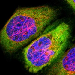
- Main image
- Experimental details
- Immunofluorescent staining of human cell line A-431 shows localization to cytosol.
- Sample type
- Human
