Explore
 Validate
 Learn
Learn Western blot
 Immunocytochemistry
Antibody data
- Antibody Data
- Antigen structure
- References [3]
- Comments [0]
- Validations
- Immunocytochemistry [2]
Submit
Validation data
Reference
Comment
Report error
- Product number
- 710209 - Provider product page

- Provider
- Invitrogen Antibodies
- Product name
- CD49f Recombinant Polyclonal Antibody (5HCLC)
- Antibody type
- Polyclonal
- Antigen
- Recombinant full-length protein
- Reactivity
- Human, Mouse
- Host
- Rabbit
- Isotype
- IgG
- Antibody clone number
- 5HCLC
- Vial size
- 100 µg
- Concentration
- 0.5 mg/mL
- Storage
- Store at 4°C short term. For long term storage, store at -20°C, avoiding freeze/thaw cycles.
Submitted references RFX1 maintains testis cord integrity by regulating the expression of Itga6 in male mouse embryos.
HPV16 infection of HaCaTs is dependent on β4 integrin, and α6 integrin processing.
Development of an in vitro 3D tumor model to study therapeutic efficiency of an anticancer drug.
Wang B, Qi T, Chen SQ, Ye L, Huang ZS, Li H
Molecular reproduction and development 2016 Jul;83(7):606-14
Molecular reproduction and development 2016 Jul;83(7):606-14
HPV16 infection of HaCaTs is dependent on β4 integrin, and α6 integrin processing.
Aksoy P, Abban CY, Kiyashka E, Qiang W, Meneses PI
Virology 2014 Jan 20;449:45-52
Virology 2014 Jan 20;449:45-52
Development of an in vitro 3D tumor model to study therapeutic efficiency of an anticancer drug.
Shin CS, Kwak B, Han B, Park K
Molecular pharmaceutics 2013 Jun 3;10(6):2167-75
Molecular pharmaceutics 2013 Jun 3;10(6):2167-75
No comments: Submit comment
Supportive validation
- Submitted by
- Invitrogen Antibodies (provider)
- Main image

- Experimental details
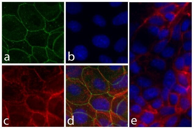
- Immunofluorescent analysis of Integrin alpha 6 in MCF-7 cells using an Integrin alpha 6 Recombinant Rabbit Polyclonal Antibody (Product # 710209) followed by detection using an Alexa Fluor 488-conjugated Goat anti-Rabbit secondary antibody (green) (Image A). Nuclei were stained using DAPI (Image B) and actin stained with Alexa Fluor 594 phalloidin (red) (image C). Image D is a composite image showing localization of integrin alpha 6 at cell junctions.
- Submitted by
- Invitrogen Antibodies (provider)
- Main image

- Experimental details
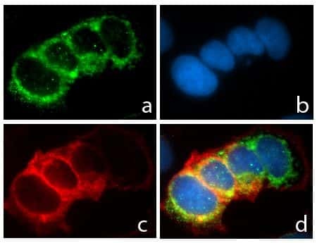
- Immunofluorescent analysis of Integrin alpha 6 was performed on 90% confluent log phase MDCK cells. The cells were fixed with 4% paraformaldehyde for 15 minutes, permeabilized with 0. 25% Triton X-100 for 10 minutes, and blocked with 5% BSA for 1 hour at room temperature. The cells were labeled with Integrin alpha 6 Recombinant Rabbit Polyclonal Antibody (Product # 710209) at a dilution of 1:500 in 1% BSA and incubated for 3 hours at room temperature and then labeled with Alexa Fluor® 488 Goat anti-Rabbit IgG secondary antibody (Product # A-11008) at a dilution of 1:400 for 30 minutes at room temperature (Panel a: green). Nuclei (Panel b: blue) were stained with SlowFade® Gold Antifade Mountant with DAPI (Product # S36938). Panel c is a merged image showing localization at the cell junctions and panel d is a control without primary antibody. The images were captured using a Nikon microscope at 20X magnification.