Explore
 Validate
 Learn
Learn Western blot
 Immunocytochemistry
Antibody data
- Antibody Data
- Antigen structure
- References [2]
- Comments [0]
- Validations
- Immunocytochemistry [5]
- Immunohistochemistry [2]
- Other assay [2]
Submit
Validation data
Reference
Comment
Report error
- Product number
- PA5-21431 - Provider product page

- Provider
- Invitrogen Antibodies
- Product name
- GANAB Polyclonal Antibody
- Antibody type
- Polyclonal
- Antigen
- Recombinant full-length protein
- Description
- Recommended positive controls: 293T, A431, H1299, HeLaS3, HepG2, Molt-4, Raji, mouse brain. Predicted reactivity: Mouse (90%), Pig (80%), Rhesus Monkey (97%), Bovine (89%). Store product as a concentrated solution. Centrifuge briefly prior to opening the vial.
- Reactivity
- Human, Mouse
- Host
- Rabbit
- Isotype
- IgG
- Vial size
- 100 μL
- Concentration
- 1 mg/mL
- Storage
- Store at 4°C short term. For long term storage, store at -20°C, avoiding freeze/thaw cycles.
Submitted references Metabolic syndrome perturbs deglucosylation and reglucosylation in the glycoprotein folding cycle.
EDEM1's mannosidase-like domain binds ERAD client proteins in a redox-sensitive manner and possesses catalytic activity.
Kuribara T, Imagawa A, Hirano M, Ito Y, Totani K
FEBS letters 2020 Jun;594(11):1759-1769
FEBS letters 2020 Jun;594(11):1759-1769
EDEM1's mannosidase-like domain binds ERAD client proteins in a redox-sensitive manner and possesses catalytic activity.
Lamriben L, Oster ME, Tamura T, Tian W, Yang Z, Clausen H, Hebert DN
The Journal of biological chemistry 2018 Sep 7;293(36):13932-13945
The Journal of biological chemistry 2018 Sep 7;293(36):13932-13945
No comments: Submit comment
Supportive validation
- Submitted by
- Invitrogen Antibodies (provider)
- Main image
- Experimental details
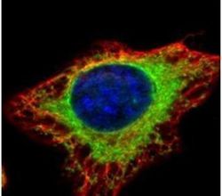
- Immunofluorescent analysis of alpha Glucosidase II in methanol-fixed HeLa cells using an Alpha Glucosidase II polyclonal antibody (Product # PA5-21431) (Green) at a 1:500 dilution. Alpha-tubulin filaments were labeled with Product # PA5-29281 (Red) at a 1:2000.
- Submitted by
- Invitrogen Antibodies (provider)
- Main image
- Experimental details
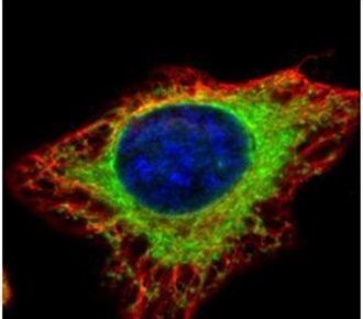
- Confocal immunofluorescence analysis (Olympus FV10i) of methanol-fixed HeLa, using alpha Glucosidase II antibody (Product # PA5-21431) (Green) at 1:500 dilution. Alpha-tubulin filaments were labeled with (Product # MA1-25054) (Red) at 1:2,000.

- Submitted by
- Invitrogen Antibodies (provider)
- Main image
- Experimental details
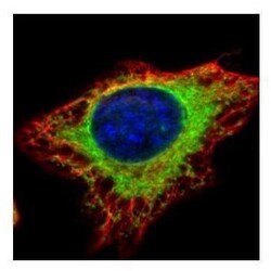
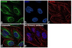
- Immunofluorescence analysis of GANAB was performed using 70% confluent log phase HeLa cells. The cells were fixed with 4% paraformaldehyde for 10 minutes, permeabilized with 0.1% Triton™ X-100 for 15 minutes, and blocked with 1% BSA for 1 hour at room temperature. The cells were labeled with GANAB Rabbit Polyclonal Antibody (Product # PA5-21431) at 5 microgram/mL in 0.1% BSA, incubated at 4 degree Celsius overnight and then labeled with Goat anti-Rabbit IgG (H+L) Superclonal™ Secondary Antibody, Alexa Fluor® 488 conjugate (Product # A27034) at a dilution of 1:2000 for 45 minutes at room temperature (Panel a: green). Nuclei (Panel b: blue) were stained with ProLong™ Diamond Antifade Mountant with DAPI (Product # P36962). F-actin (Panel c: red) was stained with Rhodamine Phalloidin (Product # R415). Panel d represents the merged image showing Cytoplasmic localization. Panel e represents control cells with no primary antibody to assess background. The images were captured at 60X magnification.

- Submitted by
- Invitrogen Antibodies (provider)
- Main image

- Experimental details
- Confocal immunofluorescence analysis (Olympus FV10i) of methanol-fixed HeLa, using alpha Glucosidase II antibody (Product # PA5-21431) (Green) at 1:500 dilution. Alpha-tubulin filaments were labeled with (Product # MA1-25054) (Red) at 1:2,000.

- Submitted by
- Invitrogen Antibodies (provider)
- Main image

- Experimental details
- Immunofluorescence analysis of GANAB was performed using 70% confluent log phase HeLa cells. The cells were fixed with 4% paraformaldehyde for 10 minutes, permeabilized with 0.1% Triton™ X-100 for 15 minutes, and blocked with 1% BSA for 1 hour at room temperature. The cells were labeled with GANAB Rabbit Polyclonal Antibody (Product # PA5-21431) at 5 microgram/mL in 0.1% BSA, incubated at 4 degree Celsius overnight and then labeled with Goat anti-Rabbit IgG (Heavy Chain) Superclonal™ Secondary Antibody, Alexa Fluor® 488 conjugate (Product # A27034) at a dilution of 1:2000 for 45 minutes at room temperature (Panel a: green). Nuclei (Panel b: blue) were stained with ProLong™ Diamond Antifade Mountant with DAPI (Product # P36962). F-actin (Panel c: red) was stained with Rhodamine Phalloidin (Product # R415). Panel d represents the merged image showing Cytoplasmic localization. Panel e represents control cells with no primary antibody to assess background. The images were captured at 60X magnification.
Supportive validation
- Submitted by
- Invitrogen Antibodies (provider)
- Main image
- Experimental details
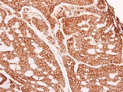
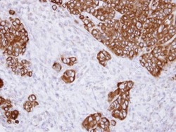
- Immunohistochemistry (Paraffin) analysis of GANAB was performed in paraffin-embedded human breast carcinoma tissue using GANAB Polyclonal Antibody (Product # PA5-21431) at a dilution of 1:500.
- Submitted by
- Invitrogen Antibodies (provider)
- Main image
- Experimental details
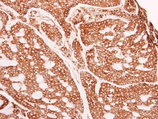
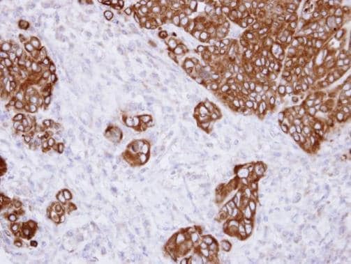
- Immunohistochemical analysis of paraffin-embedded SW480 xenograft , using alpha Glucosidase II (Product # PA5-21431) antibody at 1:500 dilution. Antigen Retrieval: Citrate buffer, pH 6.0, 15 min.
Supportive validation
- Submitted by
- Invitrogen Antibodies (provider)
- Main image
- Experimental details
- NULL
- Submitted by
- Invitrogen Antibodies (provider)
- Main image

- Experimental details
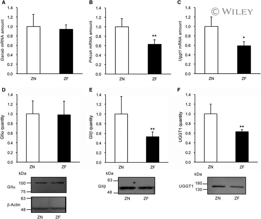
- 2 Fig. Relative amounts of glucosidase II and uridine diphosphate-glucose: glycoprotein glucosyltransferase 1 mRNA and protein in obesity model rats. Relative amounts of mRNA of Ganab (glucosidase II alpha-subunit) (A), Prkcsh (glucosidase II beta-subunit) (B), and Uggt1 (C) in Zucker normal (ZN) and Zucker fatty (ZF) rats are represented in the bar graphs. beta2 Microglobulin was used as normalization control. Relative protein quantities of glucosidase II alpha-subunit (GIIalpha) (D), glucosidase II beta-subunit (GIIbeta) (E), and uridine diphosphate-glucose: glycoprotein glucosyltransferase 1 (UGGT1) (F) in ZN and ZF rats are represented in the bar graphs. Typical western blot results are shown below the bar graphs. beta-Actin was used as loading control for normalization of all examined protein quantities. The homogenates were applied at 10 µg protein/lane. Data are expressed as mean +- standard deviation ( n = 3). Statistical significance is indicated by *, **, and *** referring to P < 0.05, 0.01, and 0.001, respectively