Explore
 Validate
 Learn
Learn Western blot
Antibody data
- Antibody Data
- Antigen structure
- References [1]
- Comments [0]
- Validations
- Western blot [5]
- Immunocytochemistry [1]
- Immunohistochemistry [2]
- Other assay [3]
Submit
Validation data
Reference
Comment
Report error
- Product number
- PA5-22144 - Provider product page

- Provider
- Invitrogen Antibodies
- Product name
- PPA1 Polyclonal Antibody
- Antibody type
- Polyclonal
- Antigen
- Recombinant full-length protein
- Description
- Recommended positive controls: H1299, HeLa, HepG2, Molt-4, Neuro 2A, GL261, C8D30, NIH-3T3, BCL-1, Raw264.7, C2C12, PC-12, Rat2. Predicted reactivity: Mouse (94%), Rat (93%), Bovine (95%). Store product as a concentrated solution. Centrifuge briefly prior to opening the vial.
- Reactivity
- Human, Mouse, Rat
- Host
- Rabbit
- Isotype
- IgG
- Vial size
- 100 μL
- Concentration
- 1.2 mg/mL
- Storage
- Store at 4°C short term. For long term storage, store at -20°C, avoiding freeze/thaw cycles.
Submitted references PPA1 regulates tumor malignant potential and clinical outcome of colon adenocarcinoma through JNK pathways.
Wang P, Zhou Y, Mei Q, Zhao J, Huang L, Fu Q
Oncotarget 2017 Aug 29;8(35):58611-58624
Oncotarget 2017 Aug 29;8(35):58611-58624
No comments: Submit comment
Supportive validation
- Submitted by
- Invitrogen Antibodies (provider)
- Main image
- Experimental details
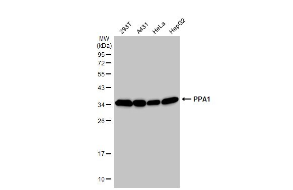
- Western Blot using PPA1 Polyclonal Antibody (Product # PA5-22144). Various whole cell extracts (30 µg) were separated by 12% SDS-PAGE, and the membrane was blotted with PPA1 Polyclonal Antibody (Product # PA5-22144) diluted at 1:10,000. The HRP-conjugated anti-rabbit IgG antibody was used to detect the primary antibody.
- Submitted by
- Invitrogen Antibodies (provider)
- Main image
- Experimental details
- PPA1 Polyclonal Antibody detects PPA1 protein by western blot analysis. A. 30 µg Neuro2A whole cell lysate/extract. B. 30 µg GL261 whole cell lysate/extract. C. 30 µg C8D30 whole cell lysate/extract. D. 30 µg NIH-3T3 whole cell lysate/extract. E. 30 µg BCL-1 whole cell lysate/extract. F. 30 µg Raw264.7 whole cell lysate/extract. G. 30 µg C2C12 whole cell lysate/extract.12% SDS-PAGE. PPA1 Polyclonal Antibody (Product # PA5-22144) dilution: 1:1,000. The HRP-conjugated anti-rabbit IgG antibody was used to detect the primary antibody.

- Submitted by
- Invitrogen Antibodies (provider)
- Main image
- Experimental details
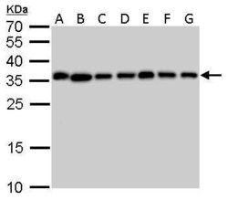
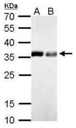
- PPA1 Polyclonal Antibody detects PPA1 protein by western blot analysis. A. 30 µg PC-12 whole cell lysate/extract. B. 30 µg Rat2 whole cell lysate/extract.12% SDS-PAGE. PPA1 Polyclonal Antibody (Product # PA5-22144) dilution: 1:1,000. The HRP-conjugated anti-rabbit IgG antibody was used to detect the primary antibody.

- Submitted by
- Invitrogen Antibodies (provider)
- Main image
- Experimental details
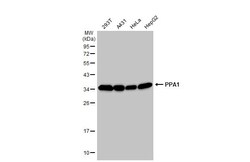
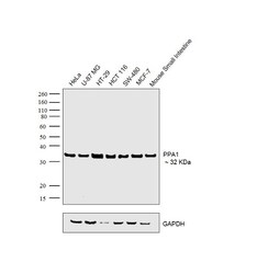
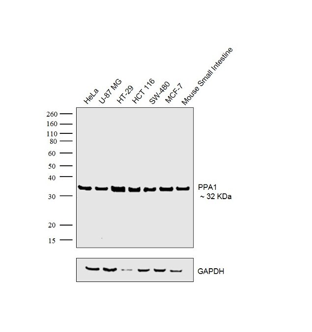
- Western blot was performed using Anti-PPA1 Rabbit Polyclonal Antibody (Product # PA5-22144) and 32 kDa band corresponding to PPA1 was observed across the samples tested. Whole cell extracts (30 µg lysate) of HeLa (Lane 1), U-87 MG (Lane 2), HT-29 (Lane 3), HCT 116 (Lane 4), SW-480 (Lane 5), MCF-7 (Lane 6) and tissue extract of Mouse Small Intestine (Lane 7) were electrophoresed using Novex® NuPAGE® 4-12% Bis-Tris gel (Product # NP0322BOX). Resolved proteins were then transferred onto a nitrocellulose membrane (Product # IB23001) by iBlot® 2 Dry Blotting System (Product # IB21001). The blot was probed with the primary antibody (1:1000 Dilution) and detected by Goat Anti-Rabbit IgG Secondary Antibody, HRP conjugate (Product # A27036, 1:4000 dilution) using the iBright FL 1000 (Product # A32752). Chemiluminescent detection was performed using Novex® ECL Chemiluminescent Substrate Reagent Kit (Product # WP20005).
- Submitted by
- Invitrogen Antibodies (provider)
- Main image

- Experimental details
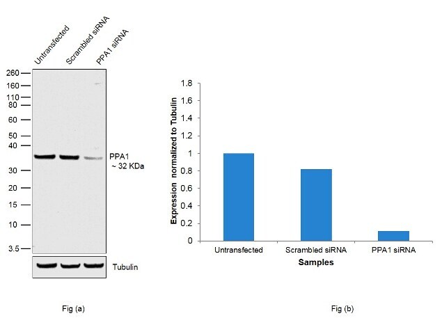
- KD of PPA1 was achieved by transfecting U-87 MG with PPA1 specific siRNAs (Silencer® select Product # s10878, s10877). Western blot analysis (Fig. a) was performed using whole cell extracts from the PPA1 KD cells (lane 3), non-specific scrambled siRNA transfected cells (lane 2) and untransfected cells (lane 1). The blots were probed with PPA1 Polyclonal Antibody (Product # PA5-22144, 1:1000 Dilution) and Goat Anti Rabbit (Heavy Chain) Superclonal™ Secondary Antibody, HRP conjugate (Product # A27036, 1:4000 dilution). Densitometric analysis of this western blot is shown in histogram (Fig. b). Decrease in signal upon siRNA mediated knock down confirms that antibody is specific to PPA1. .
Supportive validation
- Submitted by
- Invitrogen Antibodies (provider)
- Main image
- Experimental details
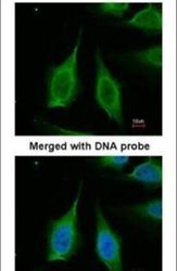
- Immunofluorescent analysis of PPA1 in paraformaldehyde-fixed HeLa cells using a PPA1 polyclonal antibody (Product # PA5-22144) at a 1:200 dilution.

Supportive validation
- Submitted by
- Invitrogen Antibodies (provider)
- Main image
- Experimental details
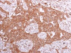
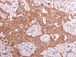
- Immunohistochemistry (Paraffin) analysis of PPA1 was performed in paraffin-embedded human breast carcinoma tissue using PPA1 Polyclonal Antibody (Product # PA5-22144) at a dilution of 1:500.
- Submitted by
- Invitrogen Antibodies (provider)
- Main image
- Experimental details
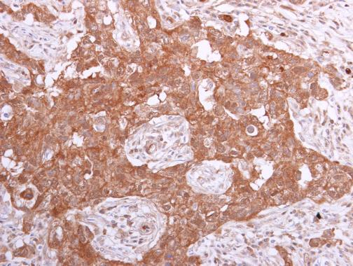
- Immunohistochemistry (Paraffin) analysis of PPA1 was performed in paraffin-embedded human breast carcinoma tissue using PPA1 Polyclonal Antibody (Product # PA5-22144) at a dilution of 1:500.

Supportive validation
- Submitted by
- Invitrogen Antibodies (provider)
- Main image
- Experimental details
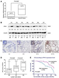
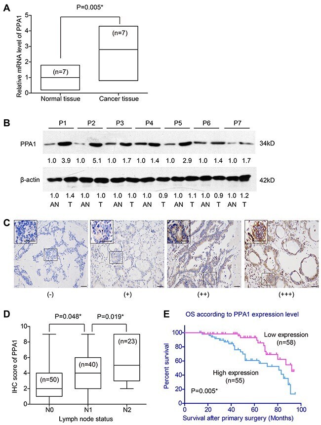
- Figure 1 Expression of PPA1 in colon adenocarcinoma tissues ( A ) RT-qPCR results showed higher mRNA levels of PPA1 in cancer tissues than that in adjacent normal tissues (P=0.005). ( B ) Western Blot revealed the different expression levels of PPA1 protein in tumor tissues (T) and adjacent normal tissues (AN). The fold changes were labelled under the bands using AN as control. P1-P7 refer to the patient's number from whom we obtained the fresh-frozen tissues. ( C ) IHC of tumor tissues showed different immunoreactivities. Scale bar: 100mum. ( D ) Patients with advanced lymph node statues exhibited higher PPA1 levels as determined by IHC evaluation. ( E ) High expression of PPA1 indicated poorer overall survival of colon adenocarcinoma patients (P=0.005).
- Submitted by
- Invitrogen Antibodies (provider)
- Main image
- Experimental details
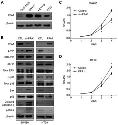
- Figure 3 PPA1 promotes proliferation in colon cancer cell lines ( A ) Western Bolt results showed that PPA1 was higher expressed in colon cancer cell lines than that in normal epithelial CCD-18Co cells. SW480 cells showed highest PPA1 expression while HT29 showed lowest PPA1 expression. ( B ) Upon PPA1-silencing, the pJNK level was significantly increased without changes in total JNK protein level. Meanwhile, PPA1 knock-down increased Bax and p53 accumulation, Bcl-2 phosphorylation as well as Caspase-3 cleavage. In contrast, overexpression of PPA1 showed reciprocal regulation towards these signaling proteins. No significant change of pERK or p-p38 levels was observed. Knock-down of PPA1 in SW480 cells inhibited cell proliferation ( C ), while overexpression of PPA1 in HT29 cells showed opposite effect ( D ). Data presented were results from three repeated experiments.

- Submitted by
- Invitrogen Antibodies (provider)
- Main image
- Experimental details
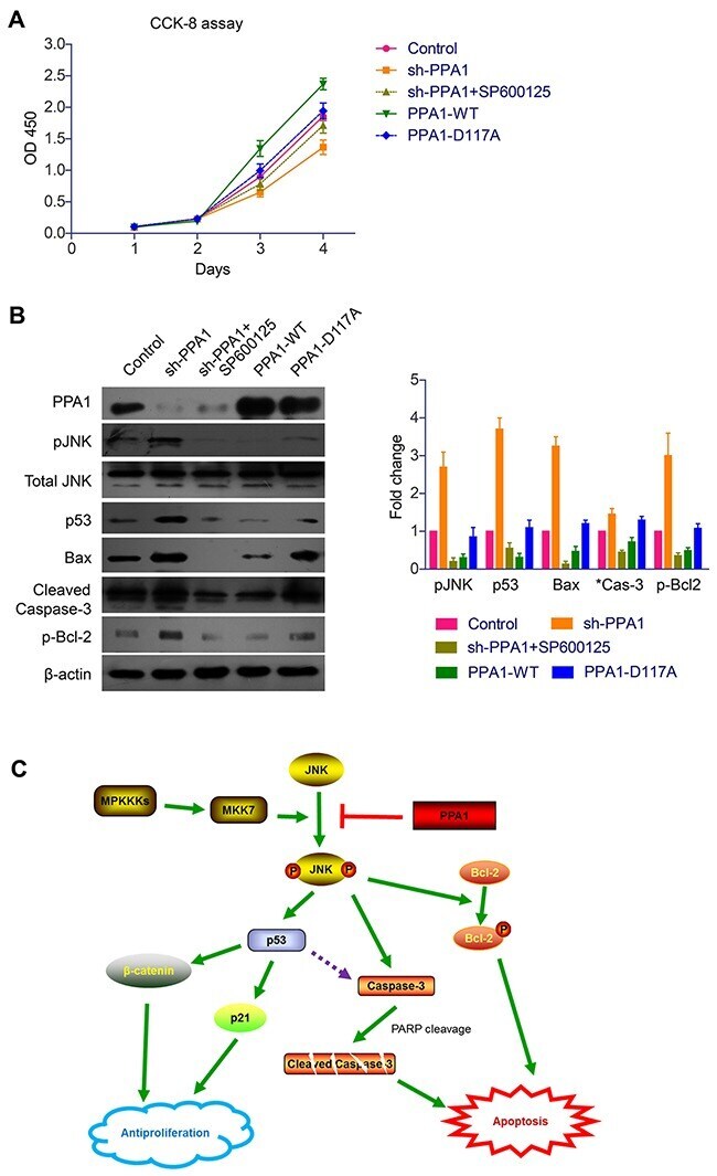
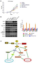
- Figure 6 PPA1 enhances cancer cell viability through inhibiting JNK signaling pathways ( A ) CCK-8 assay showed that PPA1 transfection enhanced the cell proliferation of cancer cells, while no significant effect was identified upon PPA1 inactive mutant transfection. In contrast, PPA1-silencing significantly down-regulated the cell proliferation, while JNK inhibitor can impair this antiproliferation effect. ( B ) Western Blot results demonstrated that PPA1 can modulate p53, Bax, p-Bcl-2 and cleaved Caspase-3 levels by recruiting JNK as an intermediate signaling molecular. Histogram showed the quantification results calculated by Image J Software from three experiments. ( C ) Schematically illustrations about the possible molecular and pathways involved in the PPA1 functions towards cancer transformation and development.