 Explore
 Validate
 Learn
Learn Western blot
Antibody data
- Antibody Data
- Antigen structure
- References [0]
- Comments [0]
- Validations
- Western blot [3]
- Immunohistochemistry [2]
Submit
Validation data
Reference
Comment
Report error
- Product number
- MA5-27438 - Provider product page

- Provider
- Invitrogen Antibodies
- Product name
- N-WASP Monoclonal Antibody (OTI2H5)
- Antibody type
- Monoclonal
- Antigen
- Recombinant full-length protein
- Reactivity
- Human
- Host
- Mouse
- Isotype
- IgG
- Antibody clone number
- OTI2H5
- Vial size
- 100 µL
- Concentration
- 1 mg/mL
- Storage
- -20° C, Avoid Freeze/Thaw Cycles
No comments: Submit comment
Supportive validation
- Submitted by
- Invitrogen Antibodies (provider)
- Main image
- Experimental details
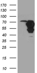
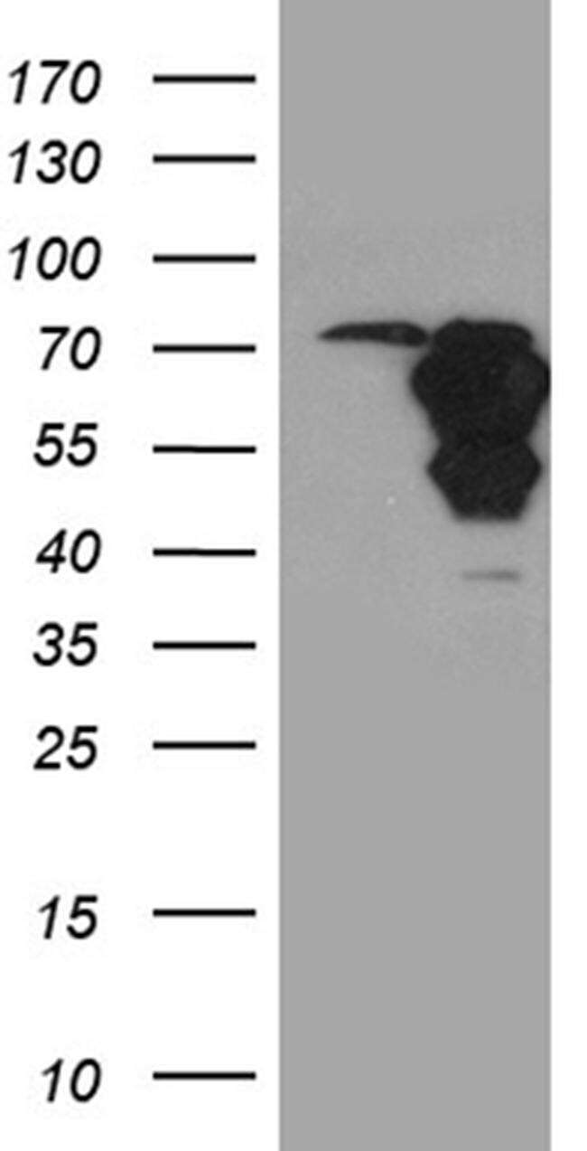
- Western blot analysis of WASL in HEK293T cells in untransfected (Left lane) and transfected (Right lane) samples using 5 µg per lane. The samples were separated by SDS-PAGE and probed with WASL (Product # MA5-27438) monoclonal antibody.
- Submitted by
- Invitrogen Antibodies (provider)
- Main image

- Experimental details
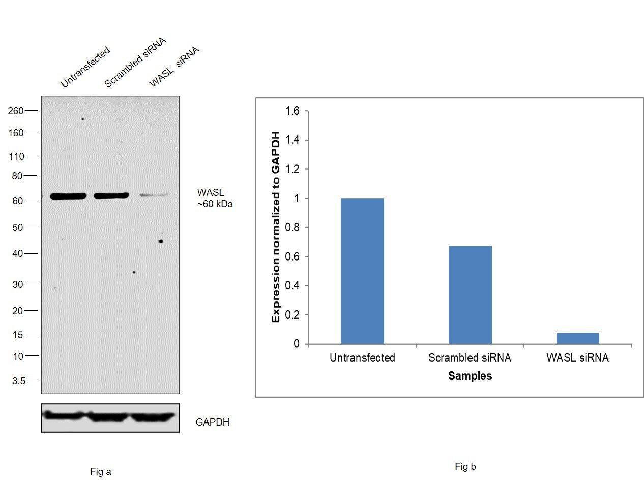
- Knockdown of N-WASP was achieved by transfecting MCF7 with N-WASP specific siRNAs (Silencer® select Product # S17133, S17132). Western blot analysis (Fig. a) was performed using Whole cell extracts from the N-WASP knockdown cells (lane 3), non-targeting scrambled siRNA transfected cells (lane 2) and untransfected cells (lane 1). The blot was probed with N-WASP Monoclonal Antibody (OTI2H5) (Product # MA5-27438, 1:2000 dilution ) and Goat anti-Mouse IgG (H+L) Superclonal™ Recombinant Secondary Antibody, HRP (Product # A28177, 1:2000 dilution). Densitometric analysis of this western blot is shown in histogram (Fig. b). Decrease in signal upon siRNA mediated knock down confirms that antibody is specific to N-WASP.
- Submitted by
- Invitrogen Antibodies (provider)
- Main image
- Experimental details
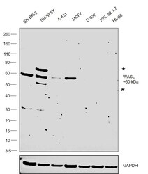
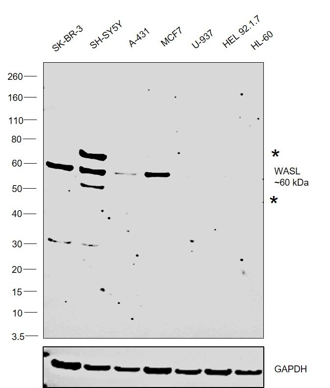
- Western blot was performed using Anti-N-WASP Monoclonal Antibody (OTI2H5) (Product # MA5-27438) and a 60 kDa band corresponding to N-WASP was observed across cell lines tested along with two uncharacterized band (*) at 50 kDa and 70 kDa. Whole cell extracts (30 µg lysate) of SK-BR-3 (Lane 1), SH-SY5Y (Lane 2), A-431 (Lane 3), MCF7 (Lane 4), U-937 (Lane 5), HEL 92.1.7 (Lane 6) and HL-60 (Lane 7) were electrophoresed using NuPAGE™ 4-12% Bis-Tris Protein Gel (Product # NP0321BOX). Resolved proteins were then transferred onto a Nitrocellulose membrane (Product # LC2001) by iBlot® 2 Dry Blotting System (Product # IB21001). The blot was probed with the primary antibody (1:2000) and detected by chemiluminescence with Goat anti-Mouse IgG (H+L) Superclonal™ Recombinant Secondary Antibody, HRP (Product # A28177, 1:4000 dilution) using the iBright FL 1000 (Product # A32752). Chemiluminescent detection was performed using Novex® ECL Chemiluminescent Substrate Reagent Kit (Product # WP20005).
Supportive validation
- Submitted by
- Invitrogen Antibodies (provider)
- Main image
- Experimental details
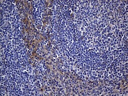
- Immunohistochemistry was performed on paraffin-embedded human tonsil tissue. To expose target proteins, heat-induced epitope retrieval by 1mM EDTA in 10mM Tris buffer (pH8.5) at 120°C for 3 min. Following antigen retrieval, tissues were probed with a WASL monoclonal antibody (Product # MA5-27438) at a dilution of 1:150.
- Submitted by
- Invitrogen Antibodies (provider)
- Main image
- Experimental details
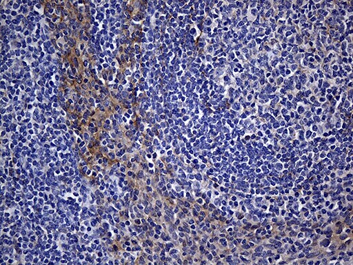
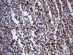
- Immunohistochemistry was performed on paraffin-embedded human spleen tissue. To expose target proteins, heat-induced epitope retrieval by 1mM EDTA in 10mM Tris buffer (pH8.5) at 120°C for 3 min. Following antigen retrieval, tissues were probed with a WASL monoclonal antibody (Product # MA5-27438) at a dilution of 1:150.
