Explore
 Validate
 Learn
Learn Western blot
 Immunocytochemistry
Antibody data
- Antibody Data
- Antigen structure
- References [9]
- Comments [0]
- Validations
- Immunocytochemistry [1]
- Immunohistochemistry [1]
Submit
Validation data
Reference
Comment
Report error
- Product number
- HPA005750 - Provider product page

- Provider
- Atlas Antibodies
- Proper citation
- Atlas Antibodies Cat#HPA005750, RRID:AB_1854729
- Product name
- Anti-WASL
- Antibody type
- Polyclonal
- Description
- Polyclonal Antibody against Human WASL, Gene description: Wiskott-Aldrich syndrome-like, Alternative Gene Names: N-WASP, NWASP, Validated applications: ICC, WB, IHC, Uniprot ID: O00401, Storage: Store at +4°C for short term storage. Long time storage is recommended at -20°C.
- Reactivity
- Human, Mouse, Rat
- Host
- Rabbit
- Conjugate
- Unconjugated
- Isotype
- IgG
- Vial size
- 100 µl
- Concentration
- 0.2 mg/ml
- Storage
- Store at +4°C for short term storage. Long time storage is recommended at -20°C.
- Handling
- The antibody solution should be gently mixed before use.
Submitted references ARPC5 isoforms and their regulation by calcium-calmodulin-N-WASP drive distinct Arp2/3-dependent actin remodeling events in CD4 T cells
Analysis of the distribution and expression of some tumor invasiveness markers in palate squamous cell carcinomas
N-Wasp Regulates Oligodendrocyte Myelination
Microridges are apical epithelial projections formed of F-actin networks that organize the glycan layer
Entry by multiple picornaviruses is dependent on a pathway that includes TNK2, WASL, and NCK1
Enteropathogenic Escherichia coli Stimulates Effector-Driven Rapid Caspase-4 Activation in Human Macrophages
Identification of the PAK4 interactome reveals PAK4 phosphorylation of N-WASP and promotion of Arp2/3-dependent actin polymerization.
N-wasp is required for stabilization of podocyte foot processes.
N-WASP coordinates the delivery and F-actin–mediated capture of MT1-MMP at invasive pseudopods
Sadhu L, Tsopoulidis N, Hasanuzzaman M, Laketa V, Way M, Fackler O
eLife 2023;12
eLife 2023;12
Analysis of the distribution and expression of some tumor invasiveness markers in palate squamous cell carcinomas
Pătru A
Romanian Journal of Morphology and Embryology 2021;61(4):1259-1278
Romanian Journal of Morphology and Embryology 2021;61(4):1259-1278
N-Wasp Regulates Oligodendrocyte Myelination
Katanov C, Novak N, Vainshtein A, Golani O, Dupree J, Peles E
The Journal of Neuroscience 2020;40(32):6103-6111
The Journal of Neuroscience 2020;40(32):6103-6111
Microridges are apical epithelial projections formed of F-actin networks that organize the glycan layer
Pinto C, Khandekar A, Bhavna R, Kiesel P, Pigino G, Sonawane M
Scientific Reports 2019;9(1)
Scientific Reports 2019;9(1)
Entry by multiple picornaviruses is dependent on a pathway that includes TNK2, WASL, and NCK1
Jiang H, Leung C, Tahan S, Wang D
eLife 2019;8
eLife 2019;8
Enteropathogenic Escherichia coli Stimulates Effector-Driven Rapid Caspase-4 Activation in Human Macrophages
Goddard P, Sanchez-Garrido J, Slater S, Kalyan M, Ruano-Gallego D, Marchès O, Fernández L, Frankel G, Shenoy A
Cell Reports 2019;27(4):1008-1017.e6
Cell Reports 2019;27(4):1008-1017.e6
Identification of the PAK4 interactome reveals PAK4 phosphorylation of N-WASP and promotion of Arp2/3-dependent actin polymerization.
Zhao M, Spiess M, Johansson HJ, Olofsson H, Hu J, Lehtiö J, Strömblad S
Oncotarget 2017 Sep 29;8(44):77061-77074
Oncotarget 2017 Sep 29;8(44):77061-77074
N-wasp is required for stabilization of podocyte foot processes.
Schell C, Baumhakl L, Salou S, Conzelmann AC, Meyer C, Helmstädter M, Wrede C, Grahammer F, Eimer S, Kerjaschki D, Walz G, Snapper S, Huber TB
Journal of the American Society of Nephrology : JASN 2013 Apr;24(5):713-21
Journal of the American Society of Nephrology : JASN 2013 Apr;24(5):713-21
N-WASP coordinates the delivery and F-actin–mediated capture of MT1-MMP at invasive pseudopods
Yu X, Zech T, McDonald L, Gonzalez E, Li A, Macpherson I, Schwarz J, Spence H, Futó K, Timpson P, Nixon C, Ma Y, Anton I, Visegrády B, Insall R, Oien K, Blyth K, Norman J, Machesky L
Journal of Cell Biology 2012;199(3):527-544
Journal of Cell Biology 2012;199(3):527-544
No comments: Submit comment
Supportive validation
- Submitted by
- Atlas Antibodies (provider)
- Main image
- Experimental details
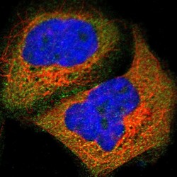
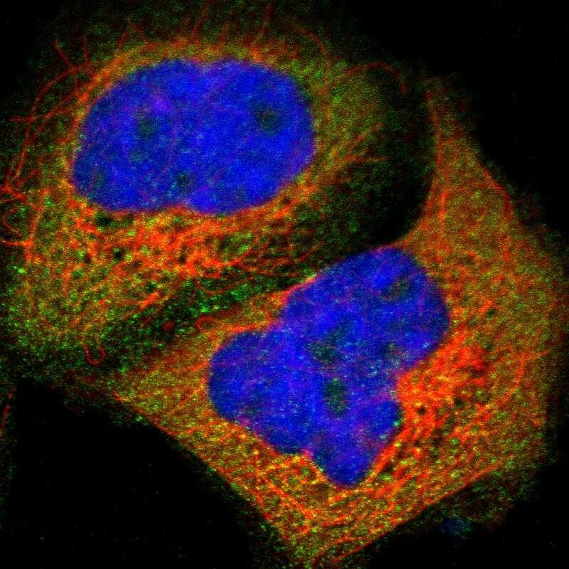
- Immunofluorescent staining of human cell line A-431 shows localization to cytosol.
- Sample type
- Human
Supportive validation
- Submitted by
- Atlas Antibodies (provider)
- Enhanced method
- Orthogonal validation
- Main image
- Experimental details
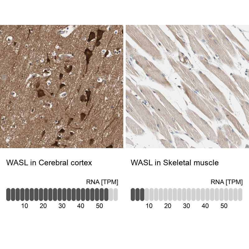
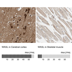
- Immunohistochemistry analysis in human cerebral cortex and skeletal muscle tissues using HPA005750 antibody. Corresponding WASL RNA-seq data are presented for the same tissues.
- Sample type
- Human
- Protocol
- Protocol