 Explore
 Validate
 Learn
Learn Western blot
Antibody data
- Antibody Data
- Antigen structure
- References [0]
- Comments [0]
- Validations
- Western blot [3]
- Immunocytochemistry [2]
Submit
Validation data
Reference
Comment
Report error
- Product number
- PA1-095 - Provider product page

- Provider
- Invitrogen Antibodies
- Product name
- KLF4 Polyclonal Antibody
- Antibody type
- Polyclonal
- Antigen
- Recombinant protein fragment
- Description
- The predicted molecular weight of KLF4 is ~55kD. The migration of this protein by SDS-PAGE was observed slightly higher (~60kD) with both PA1-095 and a benchmark antibody. PA1-095 detects a predominant band at ~60kD, and a nonspecific band of unknown origin at ~80kD in both whole cell lysates and nuclear extracts. For best results, StartingBlock T20 (TBS) Blocking Buffer (Product # 37543) is recommended.
- Reactivity
- Human, Mouse
- Host
- Rabbit
- Isotype
- IgG
- Vial size
- 200 µL
- Concentration
- 0.5 mg/mL
- Storage
- -20°C
No comments: Submit comment
Supportive validation
- Submitted by
- Invitrogen Antibodies (provider)
- Main image

- Experimental details
- Western blot analysis was performed on whole cell extracts (20 µg lysate) of HT-29 (Lane 1), HeLa (Lane 2), A-431 (Lane 3), PC-3 (lane 4), Jurkat (lane 5), HCT 116 (lane 6), K562 (lane 7), COLO 205 (lane 8), PC-12 (lane 9) and A549 (lane 10). The blots were probed with Anti-KLF4 Rabbit Polyclonal Antibody (Product # PA1-095, 1:500-1:1500 dilution) and detected by chemiluminescence using Goat anti-Rabbit IgG (H+L) Superclonal™ Secondary Antibody, HRP conjugate (Product # A27036, 0.4 µg/mL, 1:2500 dilution). A 54 kDa band corresponding to KLF4 was observed across cell lines tested expect for PC-12. Known quantity of protein samples were electrophoresed using Novex® NuPAGE® 4-12 % Bis-Tris gel (Product # NP0322BOX), XCell SureLock™ Electrophoresis System (Product # EI0002) and Novex® Sharp Pre-Stained Protein Standard (Product # LC5800). Resolved proteins were then transferred onto a nitrocellulose membrane with iBlot® 2 Dry Blotting System (Product # IB21001). The membrane was probed with the relevant primary and secondary Antibody following blocking with 5 % skimmed milk. Chemiluminescent detection was performed using Pierce™ ECL Western Blotting Substrate (Product # 32106).

- Submitted by
- Invitrogen Antibodies (provider)
- Main image
- Experimental details
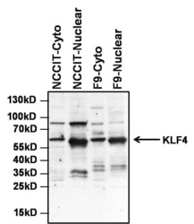
- Western blot analysis of KLF4 was performed by loading 25 µg of NCCIT and F9 cytoplasmic and nuclear extracts, and 10 µL of PageRuler Prestained Protein Ladder (Product # 26616) per well onto a 4-20% Tris-HCl polyacrylamide gel. Proteins were transferred to a PVDF membrane and blocked with StartingBlock T20 (TBS) Blocking Buffer (Product # 37543) for at least 1 hour at room temperature. The membrane was probed with a KLF4 polyclonal antibody (Product # PA1-095) at a dilution of 1:1000 overnight at 4°C on a rocking platform, washed in TBS-0.1%Tween-20, and probed with an HRP-conjugated goat anti-rabbit IgG secondary antibody (Product # 31460) at a dilution of 1:40,000 for at least 30 minutes. Chemiluminescent detection was performed using SuperSignal West Dura (Product # 34075). NOTE: Cytoplasmic and nuclear extracts were obtained using NE-PER Nuclear Protein Extraction Kit (Product # 78833).

- Submitted by
- Invitrogen Antibodies (provider)
- Main image

- Experimental details
- Western blot analysis of KLF4 was performed by loading 20 µg of HEK293T lysates from cells transfected with a control vector (left lane) or a KLF4 overexpression plasmid (right panel) and 10 µL of PageRuler Prestained Protein Ladder (Product # 26616) per well onto a 4-20% Tris-HCl polyacrylamide gel. Proteins were transferred to a PVDF membrane and blocked with 5% BSA in TBST for at least 1 hour. The membrane was probed with a KLF4 polyclonal antibody (Product # PA1-095) at a dilution of 1:1000 overnight at 4°C on a rocking platform, washed in TBS-0.1%Tween-20, and probed with an HRP-conjugated goat anti-rabbit IgG secondary antibody (Product # 31460) at a dilution of 1:40,000 for at least 30 minutes. Chemiluminescent detection was performed using SuperSignal West Pico (Product # 34080).

Supportive validation
- Submitted by
- Invitrogen Antibodies (provider)
- Main image
- Experimental details
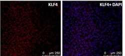
- Immunofluorescent analysis of KLF4 (red) in human embryonic stem cell H9 line grown on irradiated MEF-feeder layer. The cells were fixed with 4% paraformaldehyde at room temperature for 10 min and permeabilized with 0.25% Triton-X 100 for 5 min and blocked with the 10% BSA in PBS for 30 min at 37°C. Cells were stained with a KLF4 polyclonal antibody (Product # PA1-095) at a dilution of 1:200 in 3% BSA/PBS blocking buffer overnight at 4°C, and then incubated with a RRX-conjugated donkey anti-rabbit IgG secondary antibody at a dilution of 1:500 for 1 hour at room temperature. Nucleus DNA (blue) was stained with DAPI (Product # D1306).

- Submitted by
- Invitrogen Antibodies (provider)
- Main image

- Experimental details
- Immunofluorescence analysis of KLF4 was done on 70% confluent log phase HeLa cells. The cells were fixed with 4% paraformaldehyde for 15 minutes, permeabilized with 0.25% Triton™ X-100 for 10 minutes, and blocked with 5% BSA for 1 hour at room temperature. The cells were labeled with KLF4 Rabbit Polyclonal Antibody (Product # PA1-095) at 1 µg/mL in 1% BSA and incubated for 3 hours at room temperature and then labeled with Goat anti-Rabbit IgG (H+L) Superclonal™ Secondary Antibody, Alexa Fluor® 488 conjugate (Product # A27034) at a dilution of 1:2000 for 45 minutes at room temperature (Panel a: green). Nuclei (Panel b: blue) were stained with SlowFade® Gold Antifade Mountant with DAPI (Product # S36938). F-actin (Panel c: red) was stained with Alexa Fluor® 555 Rhodamine Phalloidin (Product # R415, 1:300). Panel d is a merged image showing nuclear localization. Panel e is a no primary antibody control. The images were captured at 60X magnification.
