Explore
 Validate
 Learn
Learn Western blot
 Immunocytochemistry
Antibody data
- Antibody Data
- Antigen structure
- References [2]
- Comments [0]
- Validations
- Immunocytochemistry [2]
- Other assay [1]
Submit
Validation data
Reference
Comment
Report error
- Product number
- PA5-27441 - Provider product page

- Provider
- Invitrogen Antibodies
- Product name
- KLF4 Polyclonal Antibody
- Antibody type
- Polyclonal
- Antigen
- Recombinant protein fragment
- Description
- Recommended positive controls: HeLa. Predicted reactivity: Mouse (89%), Rat (89%), Cat (93%), Pig (93%), Sheep (92%), Rhesus Monkey (98%), Bovine (92%). Store product as a concentrated solution. Centrifuge briefly prior to opening the vial.
- Reactivity
- Human, Mouse
- Host
- Rabbit
- Isotype
- IgG
- Vial size
- 100 µL
- Concentration
- 1.0 mg/mL
- Storage
- Store at 4°C short term. For long term storage, store at -20°C, avoiding freeze/thaw cycles.
Submitted references Inactivation of Cerebral Cavernous Malformation Genes Results in Accumulation of von Willebrand Factor and Redistribution of Weibel-Palade Bodies in Endothelial Cells.
Precise CCM1 gene correction and inactivation in patient-derived endothelial cells: Modeling Knudson's two-hit hypothesis in vitro.
Much CD, Sendtner BS, Schwefel K, Freund E, Bekeschus S, Otto O, Pagenstecher A, Felbor U, Rath M, Spiegler S
Frontiers in molecular biosciences 2021;8:622547
Frontiers in molecular biosciences 2021;8:622547
Precise CCM1 gene correction and inactivation in patient-derived endothelial cells: Modeling Knudson's two-hit hypothesis in vitro.
Spiegler S, Rath M, Much CD, Sendtner BS, Felbor U
Molecular genetics & genomic medicine 2019 Jul;7(7):e00755
Molecular genetics & genomic medicine 2019 Jul;7(7):e00755
No comments: Submit comment
Supportive validation
- Submitted by
- Invitrogen Antibodies (provider)
- Main image
- Experimental details
- KLF4 Polyclonal Antibody detects KLF4 protein at nucleus by immunofluorescent analysis. Sample: HeLa cells were fixed in 4% paraformaldehyde/PBS for 15 min. Green: KLF4 protein stained by KLF4 Polyclonal Antibody (Product # PA5-27441) diluted at 1:500. Blue: Hoechst 33342 staining.

Supportive validation
- Submitted by
- Invitrogen Antibodies (provider)
- Main image
- Experimental details
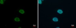
- FIGURE 2 Characterization of CCM1 +/- and clonally expanded CCM1 -/- BOECS. (A) CCM1 -/- BOECs were established by limiting dilution of the CRISPR/Cas9 RNP-treated cell pool and clonal expansion of single cells (created with BioRender.com). (B) Shown are the genotypes of CCM1 -/- BOEC clones included in this study. All variants either lead to a frameshift or a premature stop codon (additional information on the CRISPR/Cas9-induced mutations and numbers of individual clones are given in Supplementary Table S1 ). (C) CCM1 -/- BOECs presented a more compact morphology in brightfield microscopy and increased tube formation. The largest cell diameter and the number of meshes formed on Matrigel-coated plates were quantified. Scale bar indicates 400 um. Data are presented as single data points with the mean ( n = 4-6). (D) Immunofluorescence staining revealed a higher number of intercellular gaps (white arrowheads), more actin stress fibers, and a higher expression of KLF2 and KLF4 in CCM1 -/- BOECs. Representative images are shown. Scale bar indicates 100 um. Individual data points are shown with the mean ( n = 3-5). Student's t test was used for statistical analysis: * p < 0.05; ** p < 0.01; **** p < 0.0001; ns = not significant; VE-cadherin = vascular endothelial cadherin.