Explore
 Validate
 Learn
Learn Western blot
 Immunocytochemistry
Antibody data
- Antibody Data
- Antigen structure
- References [1]
- Comments [0]
- Validations
- Immunocytochemistry [1]
- Immunohistochemistry [1]
Submit
Validation data
Reference
Comment
Report error
- Product number
- AF3190 - Provider product page

- Provider
- R&D Systems
- Product name
- Mouse Syndecan-1/CD138 Antibody
- Antibody type
- Polyclonal
- Description
- Antigen Affinity-purified. Detects mouse Syndecan-1/CD138 in direct ELISAs and Western blots. In Western blots, approximately 5% cross-reactivity with recombinant human (rh) Syndecan-1 is observed and less than 1% cross-reactivity with rhSyndecan-2, recombinant mouse (rm) Syndecan-3 and rmSyndecan-4 is observed.
- Reactivity
- Mouse
- Host
- Goat
- Conjugate
- Unconjugated
- Antigen sequence
P18828- Isotype
- IgG
- Vial size
- 100 ug
- Concentration
- LYOPH
- Storage
- Use a manual defrost freezer and avoid repeated freeze-thaw cycles. 12 months from date of receipt, -20 to -70 °C as supplied. 1 month, 2 to 8 °C under sterile conditions after reconstitution. 6 months, -20 to -70 °C under sterile conditions after reconstitution.
Submitted references Heparan sulfate proteoglycans present PCSK9 to the LDL receptor.
Gustafsen C, Olsen D, Vilstrup J, Lund S, Reinhardt A, Wellner N, Larsen T, Andersen CBF, Weyer K, Li JP, Seeberger PH, Thirup S, Madsen P, Glerup S
Nature communications 2017 Sep 11;8(1):503
Nature communications 2017 Sep 11;8(1):503
No comments: Submit comment
Supportive validation
- Submitted by
- R&D Systems (provider)
- Main image
- Experimental details
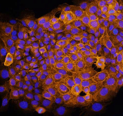
- Syndecan-1/CD138 in NMuMG Mouse Cell Line. Syndecan-1/CD138 was detected in immersion fixed NMuMG mouse mammary gland epithelial cell line using Mouse Syndecan-1/CD138 Antigen Affinity-purified Polyclonal Antibody (Catalog # AF3190) at 10 µg/mL for 3 hours at room temperature. Cells were stained using the NorthernLights™ 557-conjugated Anti-Goat IgG Secondary Antibody (yellow; Catalog # NL001) and counterstained with DAPI (blue). Specific staining was localized to cell surfaces and cytoplasm. View our protocol for Fluorescent ICC Staining of Cells on Coverslips.

Supportive validation
- Submitted by
- R&D Systems (provider)
- Main image

- Experimental details
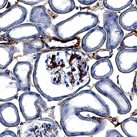
- Syndecan-1/CD138 in Mouse Kidney. Syndecan-1/CD138 was detected in perfusion fixed frozen sections of mouse kidney using Goat Anti-Mouse Syndecan-1/CD138 Antigen Affinity-purified Polyclonal Antibody (Catalog # AF3190) at 3 µg/mL for 1 hour at room temperature followed by incubation with the Anti-Goat IgG VisUCyte™ HRP Polymer Antibody (Catalog # VC004). Tissue was stained using DAB (brown) and counterstained with hematoxylin (blue). Specific staining was localized to glomeruli and convoluted tubules. View our protocol for IHC Staining with VisUCyte HRP Polymer Detection Reagents.