 Explore
 Validate
 Learn
Learn Western blot
 Immunohistochemistry
Antibody data
- Antibody Data
- Antigen structure
- References [1]
- Comments [0]
- Validations
- Immunohistochemistry [2]
- Other assay [2]
Submit
Validation data
Reference
Comment
Report error
- Product number
- PA5-47395 - Provider product page

- Provider
- Invitrogen Antibodies
- Product name
- CD138 Polyclonal Antibody
- Antibody type
- Polyclonal
- Antigen
- Recombinant full-length protein
- Description
- In sandwich ELISAs, less than 0.3% cross-reactivity with recombinant human (rh) Syndecan-2, rhSyndecan-3, rhSyndecan-4, and recombinant mouse Syndecan-1 is observed. Reconstitute at 0.2 mg/mL in sterile PBS.
- Reactivity
- Human
- Host
- Goat
- Isotype
- IgG
- Vial size
- 100 µg
- Concentration
- 0.2 mg/mL
- Storage
- -20° C, Avoid Freeze/Thaw Cycles
Submitted references Ectopic Lymphoid Follicle Formation and Human Seasonal Influenza Vaccination Responses Recapitulated in an Organ-on-a-Chip.
Goyal G, Prabhala P, Mahajan G, Bausk B, Gilboa T, Xie L, Zhai Y, Lazarovits R, Mansour A, Kim MS, Patil A, Curran D, Long JM, Sharma S, Junaid A, Cohen L, Ferrante TC, Levy O, Prantil-Baun R, Walt DR, Ingber DE
Advanced science (Weinheim, Baden-Wurttemberg, Germany) 2022 May;9(14):e2103241
Advanced science (Weinheim, Baden-Wurttemberg, Germany) 2022 May;9(14):e2103241
No comments: Submit comment
Supportive validation
- Submitted by
- Invitrogen Antibodies (provider)
- Main image
- Experimental details
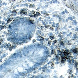
- Immunohistochemical analysis of Syndecan-1/CD138 was detected in immersion fixed paraffin-embedded sections of human ileum using human Syndecan-1/CD138 Antigen Affinity-purified Polyclonal Antibody (Product # PA5-47395) at 15 µg/mL overnight at 4 °C. Tissue was stained using the Anti-Goat HRP-DAB (brown and counterstained with hematoxylin (blue).

- Submitted by
- Invitrogen Antibodies (provider)
- Main image

- Experimental details
- Immunohistochemical analysis of Syndecan-1/CD138 was detected in immersion fixed paraffin-embedded sections of human jejunum using human Syndecan-1/CD138 Antigen Affinity-purified Polyclonal Antibody (Product # PA5-47395) at 15 µg/mL overnight at 4 °C. Tissue was stained using the Anti-Goat HRP-DAB (brown and counterstained with hematoxylin (blue). Lower panel shows a lack of labeling if primary antibodies are omitted and tissue is stained only with secondary antibody followed by incubation with detection reagents.

Supportive validation
- Submitted by
- Invitrogen Antibodies (provider)
- Main image

- Experimental details
- Influenza vaccination in vitro in the human LF chip. a) Violin plot of anti-HA IgG levels that are specific to the Brisbane 59 H1N1 strain (Anti-HA IgG) in the effluent of LF Chips 9 days after vaccination with Fluzone presented relative to levels measured in a vaccinated culture of tonsillar cells (Tonsillar Control), as detected using a digital ELISA. Each data point represents average of 2-6 chips created with cells from one donor (total 8 donors tested). b) Time course of anti-HA IgG secretion measured in chip effluents over 5 to 12 days of culture using LF Chips containing cells from representative donors from the high () and low (V) Ab producer groups shown in a. c) Immunofluorescence micrograph showing CD138 staining (green) in a Fluzone-stimulated LF Chip containing cells (nuclei, magenta) from a high anti-HA Ab producer (similar results are obtained with 3 different high Ab producer donors; bar, 100 um). d) Anti-HA Ab levels that are specific to the Brisbane 59 H1N1 strain in the effluent of LF Chips with or without DCs, 9 days after vaccination, as detected by a digital ELISA, presented relative to levels measured in a culture of tonsillar cells. Mean levels from 3 replicate measurements from one chip generated from one donor are shown, and similar results are obtained in LF Chips created with cells from two different donors. Error bars indicate SD; *, p < 0.05 using an unpaired Student's t-test with Welch's correction. e) CXCL13 levels in the effluent of the LF chi

- Submitted by
- Invitrogen Antibodies (provider)
- Main image

- Experimental details
- Figure 3 B cells exhibit class switching and undergo plasma cell formation in the LF chip. a) Total IgG production measured in the effluents of LF chips when engineered with naive B cells and bulk T cells after 6 days culture in the presence or absence (-) of IL4 and anti-CD40 Ab Each dot indicates results from an individual chip ( n = 2) created with cells from two donors (black and gray); *, p < 0.05 using an unpaired Student's t -test with Welch's correction. b) Immunofluorescence micrographs showing cells in unstimulated LF chips (No stim.) or chips treated with IL4 and anti-CD40 Ab stained for CD138 (green) and nuclei (magenta); similar results are obtained with cells from 3 different donors. c) Quantification of CD138 expression in single cells (gray bars) versus cells located within LFs (black bars) in the same LF chips. Error bars indicate SD based on analysis of 5 randomly selected fields from 1 donor, and similar results are obtained with LF Chips containing cells from 3 different donors. *, p < 0.05 using a two way ANOVA to identify any significant differences followed by the Fisher's LSD test. d) Immunostaining for CD138 (green) and nuclei (magenta) in SAC-treated LF Chips (similar results obtained with 3 donors; bar 100 um). e) CD138 levels measured as a % of projected area labeled for CD138 in lone cells (Single Cells) versus cells in follicles (LFs) within ECM gels in perfused Organ Chips. Results shown are from 5 randomly selected fields from 1 LF Chip created
