 Explore
 Validate
 Learn
Learn Immunocytochemistry
Antibody data
- Antibody Data
- Antigen structure
- References [0]
- Comments [0]
- Validations
- Immunocytochemistry [1]
- Immunohistochemistry [1]
- Flow cytometry [1]
- Other assay [1]
Submit
Validation data
Reference
Comment
Report error
- Product number
- MAB2780 - Provider product page

- Provider
- R&D Systems
- Product name
- Human Syndecan-1/CD138 Antibody
- Antibody type
- Monoclonal
- Description
- Protein A or G purified from hybridoma culture supernatant. Detects human Syndecan-1 in direct ELISAs. In direct ELISAs, this antibody shows approximately 60% cross-reactivity with recombinant mouse (rm) Syndecan-1 and no cross-reactivity with recombinant human (rh) Syndecan-2, rhSyndecan-3, or rmSyndecan-4.
- Reactivity
- Human
- Host
- Rat
- Conjugate
- Unconjugated
- Antigen sequence
NP_002988- Isotype
- IgG
- Antibody clone number
- 359103
- Vial size
- 100 ug
- Concentration
- LYOPH
- Storage
- Use a manual defrost freezer and avoid repeated freeze-thaw cycles. 12 months from date of receipt, -20 to -70 °C as supplied. 1 month, 2 to 8 °C under sterile conditions after reconstitution. 6 months, -20 to -70 °C under sterile conditions after reconstitution.
No comments: Submit comment
Supportive validation
- Submitted by
- R&D Systems (provider)
- Main image
- Experimental details
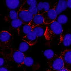
- Syndecan-1/CD138 in U266 Human Cell Line. Syndecan-1/CD138 was detected in immersion fixed U266 human myeloma cell line using Rat Anti-Human Syndecan-1/CD138 Monoclonal Antibody (Catalog # MAB2780) at 3 µg/mL for 3 hours at room temperature. Cells were stained using the NorthernLights™ 557-conjugated Anti-Rat IgG Secondary Antibody (red; Catalog # NL013) and counterstained with DAPI (blue). Specific staining was localized to plasma membrane. View our protocol for Fluorescent ICC Staining of Non-adherent Cells.

Supportive validation
- Submitted by
- R&D Systems (provider)
- Main image

- Experimental details
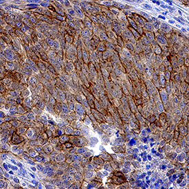
- Syndecan-1/CD138 in Human Cervical Cancer Tissue. Syndecan-1/CD138 was detected in immersion fixed paraffin-embedded sections of human cervical cancer tissue using Rat Anti-Human Syndecan-1/CD138 Monoclonal Antibody (Catalog # MAB2780) at 1.7 µg/mL overnight at 4 °C. Tissue was stained using the Anti-Rat HRP-DAB Cell & Tissue Staining Kit (brown; Catalog # CTS017) and counterstained with hematoxylin (blue). Specific staining was localized to plasma membrane. View our protocol for Chromogenic IHC Staining of Paraffin-embedded Tissue Sections.
Supportive validation
- Submitted by
- R&D Systems (provider)
- Main image
- Experimental details
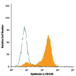
- Detection of Syndecan-1/CD138 in RPMI 8226 Human Cell Line by Flow Cytometry. RPMI 8226 human multiple myeloma cell line was stained with Rat Anti-Human Syndecan-1/CD138 Monoclonal Antibody (Catalog # MAB2780, filled histogram) or isotype control antibody (Catalog # MAB005, open histogram), followed by Phycoerythrin-conjugated Anti-Rat IgG Secondary Antibody (Catalog # F0105B).

Supportive validation
- Submitted by
- R&D Systems (provider)
- Main image
- Experimental details
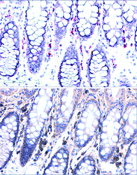
- Syndecan-1/CD138 in Human Colon Using Dual RNAscope® ISH and IHC. Syndecan-1/CD138 mRNA was detected in formalin-fixed paraffin-embedded tissue sections of human colon probed with ACD RNAScope® Probe (Catalog # 416961) and stained using ACD RNAscope® 2.5 HD Duplex Detection Reagents (red, Catalog # 322500). Adjacent tissue section was processed for immunohistochemistry using R&D Systems Rat Anti-Human Syndecan-1/CD138 Monoclonal Antibody (Catalog# MAB2780) at 0.5 ug/mL for 1 hour at room temperature followed by incubation with the Anti-Rat IgG VisUCyte HRP Polymer Antibody (R&D Systems, Catalog # VC005) and DAB chromogen (brown). Tissues were counterstained with hematoxylin (blue).
