Explore
 Validate
 Learn
Learn Western blot
 Immunohistochemistry
Antibody data
- Antibody Data
- Antigen structure
- References [0]
- Comments [0]
- Validations
- Western blot [2]
- Immunocytochemistry [2]
Submit
Validation data
Reference
Comment
Report error
- Product number
- HPA024089 - Provider product page

- Provider
- Atlas Antibodies
- Proper citation
- Atlas Antibodies Cat#HPA024089, RRID:AB_1848278
- Product name
- Anti-ETFA
- Antibody type
- Polyclonal
- Description
- Polyclonal Antibody against Human ETFA, Gene description: electron-transfer-flavoprotein, alpha polypeptide, Alternative Gene Names: EMA, GA2, MADD, Validated applications: ICC, IHC, WB, Uniprot ID: P13804, Storage: Store at +4°C for short term storage. Long time storage is recommended at -20°C.
- Reactivity
- Human, Mouse, Rat
- Host
- Rabbit
- Conjugate
- Unconjugated
- Isotype
- IgG
- Vial size
- 100 µl
- Concentration
- 0.2 mg/ml
- Storage
- Store at +4°C for short term storage. Long time storage is recommended at -20°C.
- Handling
- The antibody solution should be gently mixed before use.
No comments: Submit comment
Enhanced validation
Enhanced validation
- Submitted by
- klas2
- Enhanced method
- Genetic validation
- Main image
- Experimental details
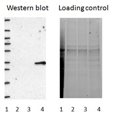
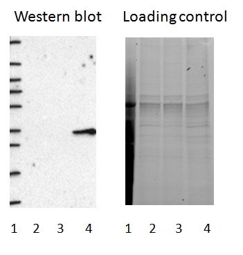
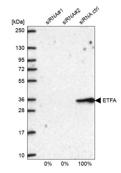
- Western blot of cell lysate from U-2 OS cells transfected with either siRNA targeting ETFA or control siRNA. Lane 1: Marker (250, 130, 95, 72, 55, 36, 28, 17, 10) Lane 2: Cell lysate from U-2OS cells transfected with siRNA targeting ETFA Lane 3: N/A Lane 4: Cell lysate from U-2OS cells transfected with control siRNA Right image, lane 1-4: loading control
- Sample type
- U-2 OS
- Primary Ab dilution
- 1:261
- Conjugate
- Horseradish Peroxidase
- Secondary Ab
- Secondary Ab
- Secondary Ab dilution
- 1:3000
- Knockdown/Genetic Approaches Application
- Western blot
Enhanced validation
- Submitted by
- Atlas Antibodies (provider)
- Enhanced method
- Genetic validation
- Main image
- Experimental details
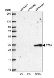
- Western blot analysis in U2OS cells transfected with control siRNA, target specific siRNA probe #1 and #2, using Anti-ETFA antibody. Remaining relative intensity is presented.
- Sample type
- Human
- Protocol
- Protocol
Enhanced validation
Supportive validation
- Submitted by
- 55af80e3e0991
- Enhanced method
- Genetic validation
- Main image
- Experimental details
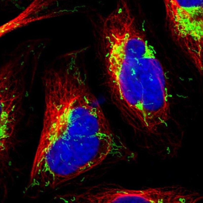
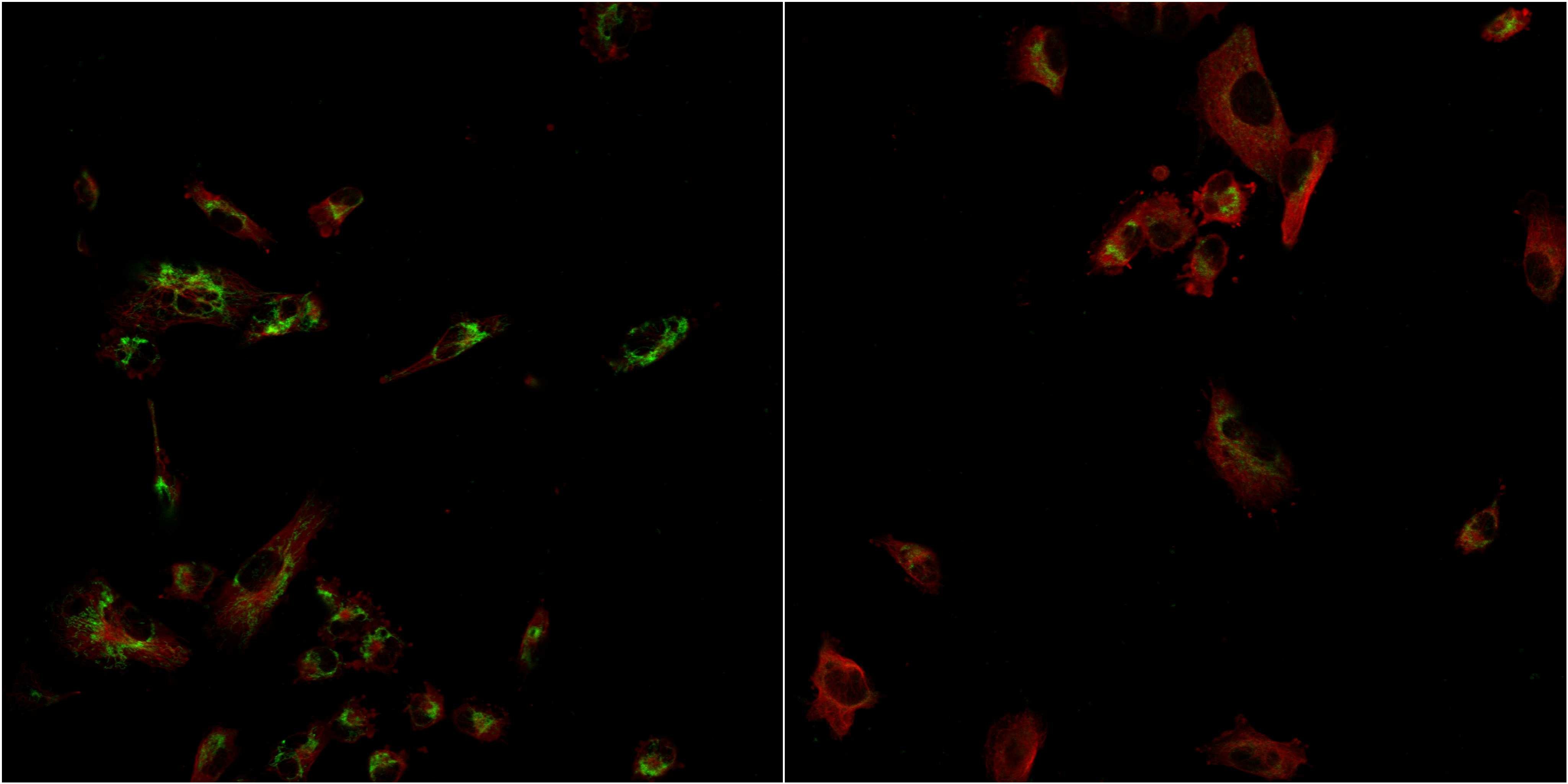
- Confocal images of immunofluorescently stained human U-2 OS cells.The protein ETFA is shown in green and the microtubules in red. The image to the left show cells transfected with control siRNA and the image to the right show cells where ETFA has been downregulated with specific siRNA.
- Sample type
- U-2 OS cells
- Primary Ab dilution
- 1:118
- Secondary Ab
- Secondary Ab
- Secondary Ab dilution
- 1:800
- Knockdown/Genetic Approaches Application
- Immunocytochemistry
Supportive validation
- Submitted by
- Atlas Antibodies (provider)
- Main image

- Experimental details
- Immunofluorescent staining of human cell line U-2 OS shows localization to mitochondria.
- Sample type
- Human