Explore
 Validate
 Learn
Learn Western blot
 ELISA
ELISAAntibody data
- Antibody Data
- Antigen structure
- References [0]
- Comments [0]
- Validations
- Western blot [2]
- Immunocytochemistry [1]
- Immunohistochemistry [2]
Submit
Validation data
Reference
Comment
Report error
- Product number
- 600-101-ML2 - Provider product page

- Provider
- Invitrogen Antibodies
- Product name
- PI3 Kinase p55 gamma Polyclonal Antibody
- Antibody type
- Polyclonal
- Antigen
- Synthetic peptide
- Reactivity
- Human, Mouse
- Host
- Goat
- Isotype
- IgG
- Vial size
- 100 µg
- Concentration
- 1.02 mg/mL
- Storage
- -20° C, Avoid Freeze/Thaw Cycles
No comments: Submit comment
Supportive validation
- Submitted by
- Invitrogen Antibodies (provider)
- Main image
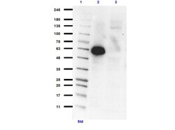
- Experimental details
- Western Blot of Goat Anti-PIK3R3 Antibody. Lane 1: Opal PreStained Molecular Weight Marker (p/n MB-210-0500) . Lane 2: PIK3R3 overexpressing lysate HEK293T lysate [+]. Lane 3: HEK293T Empty vector lysate [-]. Primary Antibody: Anti-PI 3 Kinase p55 gamma antibody at 1:1000 overnight at 2-8°C. Secondary Antibody: Donkey Anti-Goat IgG HRP (p/n 605-703-002) at 1:40,000 for 30 minutes at RT. Block: BlockOut Buffer (p/n MB-073). Predicted MW: ~54kDa overexpressed. ~44, 47, 54kDa endogenous human. Observed MW: ~57-65kDa. Exposure: 75 sec.

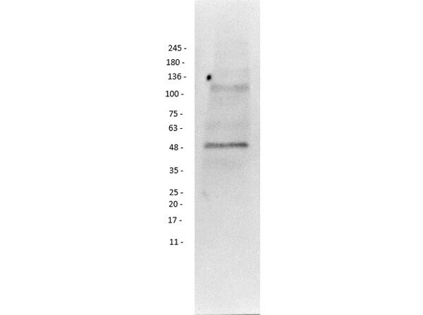
- Submitted by
- Invitrogen Antibodies (provider)
- Main image

- Experimental details
- Western Blot of Goat Anti-PIK3R3 Antibody. Lane 1: Mouse Testes Lysate. Primary Antibody: Anti-PI 3 Kinase p55 gamma antibody at 1:1000 overnight at 2-8°C. Secondary Antibody: Donkey Anti-Goat IgG HRP (p/n 605-703-125) at 1:5000 for 30 minutes at RT. Block: BlockOut Buffer (p/n MB-073). Predicted MW: ~54kDa mouse. Observed MW: ~50kDa. Exposure: 1 sec.
Supportive validation
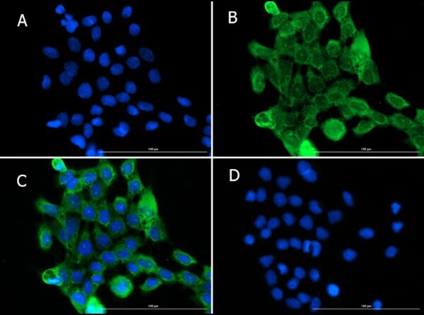
- Submitted by
- Invitrogen Antibodies (provider)
- Main image

- Experimental details
- Immunofluorescence of Goat Anti-PIK3R3 Antibody. Cell Line: A431 Cells. Fixation: 100% MeOH. Permeabilization: 0.3% Triton X-100. Primary Antibody: Anti-PI 3 Kinase p55 gamma Antibody at 15µg/mL overnight at 2-8°C. Secondary Antibody: Donkey Anti-Goat IgG DyLight™488 (p/n 605-741-125) at 15µg/mL for 1hr at RT. Nuclear Counterstain: DAPI. Localization: Cytosol (UniProtKB), Nucleoplasms (HPA) distinct punctate staining in nucleus could indicate nucleoplasm. Staining: (A) DAPI, (B) Anti-PIK3R3 + secondary, (C) A+B merge, (D) secondary only.
Supportive validation
- Submitted by
- Invitrogen Antibodies (provider)
- Main image
- Experimental details
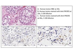
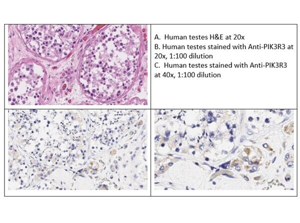
- Immunohistochemistry of Goat anti-PIK3R3 Antibody. Tissue: Human Testes. Fixative: none. Antigen Retrieval: HIER using Citrate buffer for 20 minutes. Primary Antibody: Anti-PI 3 Kinase p55 gamma antibody at 1:100 for 30 minutes at RT. Secondary Antibody: Ready-to-Use Donkey anti-Goat HRP. Counterstain: Hematoxylin. Substrate: DAB. Results: PIK3R3 shows diffuse weak cytoplasmic expression in human testis for all germ cells and Leydig cells.
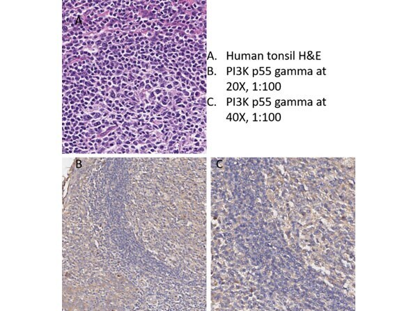
- Submitted by
- Invitrogen Antibodies (provider)
- Main image
- Experimental details
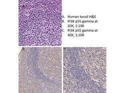
- Immunohistochemistry of Goat anti-PIK3R3 Antibody. Tissue: Human Tonsil. Fixative: none. Antigen Retrieval: HIER using Citrate buffer for 20 minutes. Primary Antibody: Anti-PI 3 Kinase p55 gamma antibody at 1:100 for 30 minutes at RT. Secondary Antibody: Donkey anti-Goat HRP at 4µL/mL for 2 minutes at RT. Counterstain: Hematoxylin. Substrate: DAB. Results: PIK3R3 showed universal tonsillar staining with increased cytoplasmic expression in germinal centers.