Explore
 Validate
 Learn
Learn Western blot
 Immunoprecipitation
Antibody data
- Antibody Data
- Antigen structure
- References [0]
- Comments [0]
- Validations
- Western blot [3]
- Immunocytochemistry [7]
- Chromatin Immunoprecipitation [2]
Submit
Validation data
Reference
Comment
Report error
- Product number
- MA5-14792 - Provider product page

- Provider
- Invitrogen Antibodies
- Product name
- JMJD1B Monoclonal Antibody (S.393.0)
- Antibody type
- Monoclonal
- Antigen
- Synthetic peptide
- Description
- It is not recommended to aliquot this antibody.
- Reactivity
- Human
- Host
- Rabbit
- Isotype
- IgG
- Antibody clone number
- S.393.0
- Vial size
- 100 μL
- Concentration
- 49 μg/mL
- Storage
- -20°C
No comments: Submit comment
Supportive validation
- Submitted by
- Invitrogen Antibodies (provider)
- Main image
- Experimental details
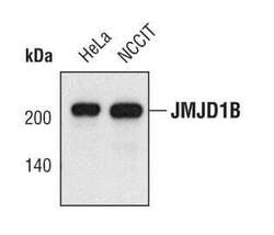
- Western blot analysis of JMJD1B in extracts from HeLa and NCCIT cell lines using JMJD1B monoclonal antibody (Product # MA5-14792).

- Submitted by
- Invitrogen Antibodies (provider)
- Main image
- Experimental details
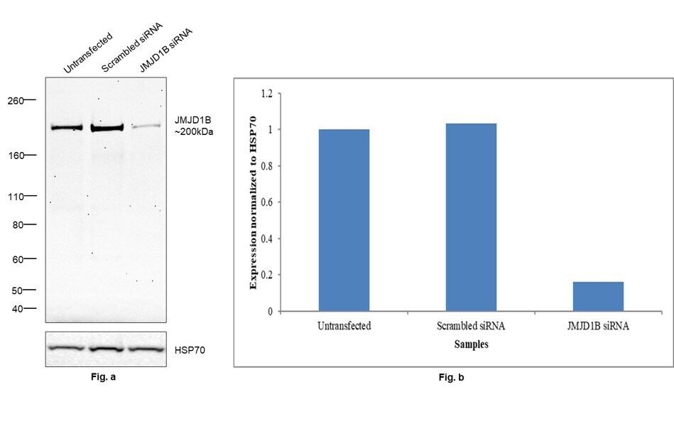
- Knockdown of JMJD1B was achieved by transfecting IMR-32 with JMJD1B specific siRNAs (Silencer® select Product # s28657, s224243). Western blot analysis (Fig. a) was performed using nuclear enriched extracts from the JMJD1B knockdown cells (lane 3), non-targeting scrambled siRNA transfected cells (lane 2) and untransfected cells (lane 1). The blot was probed with JMJD1B Monoclonal Antibody (S.393.0) (Product # MA5-14792, 1:1000 dilution ) and Goat anti-Rabbit IgG (Heavy Chain) Superclonal™ Recombinant Secondary Antibody, HRP (Product # A27036, 1:20000 dilution). Densitometric analysis of this western blot is shown in histogram (Fig. b). Decrease in signal upon siRNA mediated knock down confirms that antibody is specific to JMJD1B.
- Submitted by
- Invitrogen Antibodies (provider)
- Main image
- Experimental details
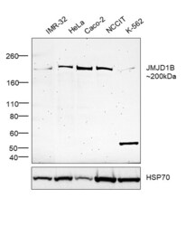
- Western blot was performed using Anti-JMJD1B Monoclonal Antibody (S.393.0) (Product # MA5-14792) and a 200 kDa band corresponding to JMJD1B was observed across all cell lines tested. Nuclear enriched extracts (50 µg lysate) of IMR-32 (Lane 1), HeLa (Lane 2), Caco-2 (Lane 3), NCCIT (Lane 4) and K-562 (Lane 5) were electrophoresed using NuPAGE™ 3-8% Tris-Acetate Protein Gel (Product # EA0378BOX). Resolved proteins were then transferred onto a nitrocellulose membrane (Product # IB23002) by iBlot® 2 Dry Blotting System (Product # IB21001). The blot was probed with the primary antibody (1:1000 dilution) and detected by chemiluminescence with Goat anti-Rabbit IgG (Heavy Chain) Superclonal™ Recombinant Secondary Antibody, HRP (Product # A27036,1:20000 dilution) using the iBright™ FL1500 Imaging System (Product # A44115). Chemiluminescent detection was performed using SuperSignal™ West Pico PLUS Chemiluminescent Substrate (Product # 34580).

Supportive validation
- Submitted by
- Invitrogen Antibodies (provider)
- Main image
- Experimental details
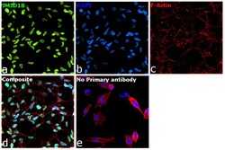
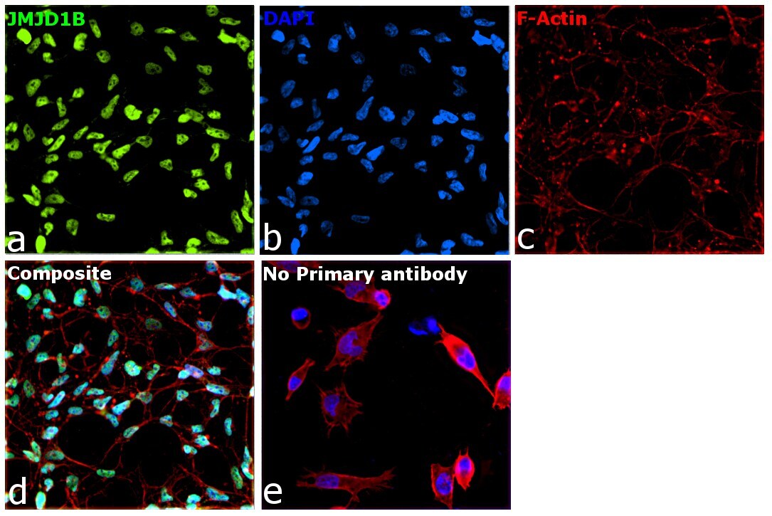
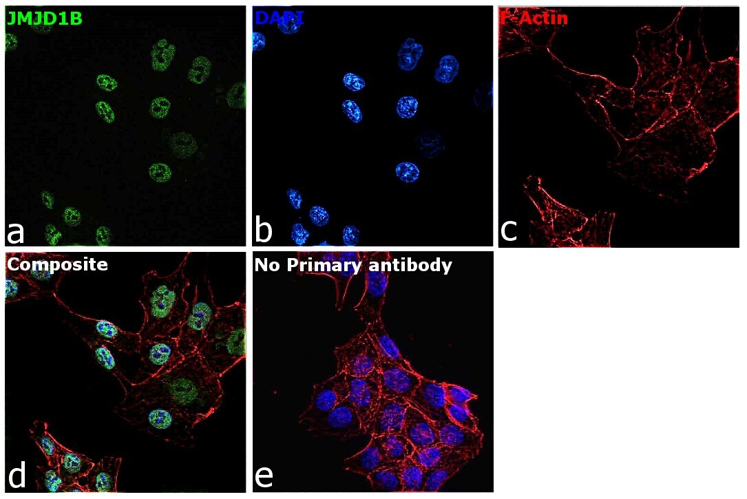
- Immunofluorescence analysis of JMJD1B was performed using 70% confluent log phase IMR-32 cells. The cells were fixed with 4% paraformaldehyde for 10 minutes, permeabilized with 0.1% Triton™ X-100 for 15 minutes, and blocked with 2% BSA for 1 hour at room temperature. The cells were labeled with JMJD1B Monoclonal Antibody (S.393.0) (Product # MA5-14792) at 1:200 dilution in 0.1% BSA, incubated at 4 degree celsius overnight and then labeled with Donkey anti-Rabbit IgG (H+L) Highly Cross-Adsorbed Secondary Antibody, Alexa Fluor Plus 488 (Product # A32790), (1:2000 dilution), for 45 minutes at room temperature (Panel a: Green). Nuclei (Panel b:Blue) were stained with ProLong™ Diamond Antifade Mountant with DAPI (Product # P36962). F-actin (Panel c: Red) was stained with Rhodamine Phalloidin (Product # R415, 1:300). Panel d represents the merged image showing nuclear localization. Panel e represents control cells with no primary antibody to assess background. The images were captured at 60x magnification.
- Submitted by
- Invitrogen Antibodies (provider)
- Main image

- Experimental details
- Immunofluorescence analysis of JMJD1B was performed using 70% confluent log phase HCT 116 cells. The cells were fixed with 4% paraformaldehyde for 10 minutes, permeabilized with 0.1% Triton™ X-100 for 15 minutes, and blocked with 2% BSA for 1 hour at room temperature. The cells were labeled with JMJD1B Monoclonal Antibody (S.393.0) (Product # MA5-14792) at 1:100 dilution in 0.1% BSA, incubated at 4 degree celsius overnight and then labeled with Goat anti-Rabbit IgG (Heavy Chain) Superclonal™ Recombinant Secondary Antibody, Alexa Fluor® 488 conjugate (Product # A27034), (1:2000 dilution), for 45 minutes at room temperature (Panel a: Green). Nuclei (Panel b:Blue) were stained with ProLong™ Diamond Antifade Mountant with DAPI (Product # P36962). F-actin (Panel c: Red) was stained with Rhodamine Phalloidin (Product # R415, 1:300). Panel d represents the merged image showing nuclear localization. Panel e represents control cells with no primary antibody to assess background. The images were captured at 60X magnification.

- Submitted by
- Invitrogen Antibodies (provider)
- Main image
- Experimental details
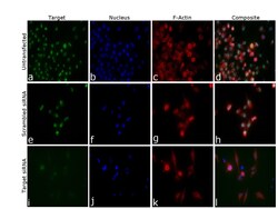
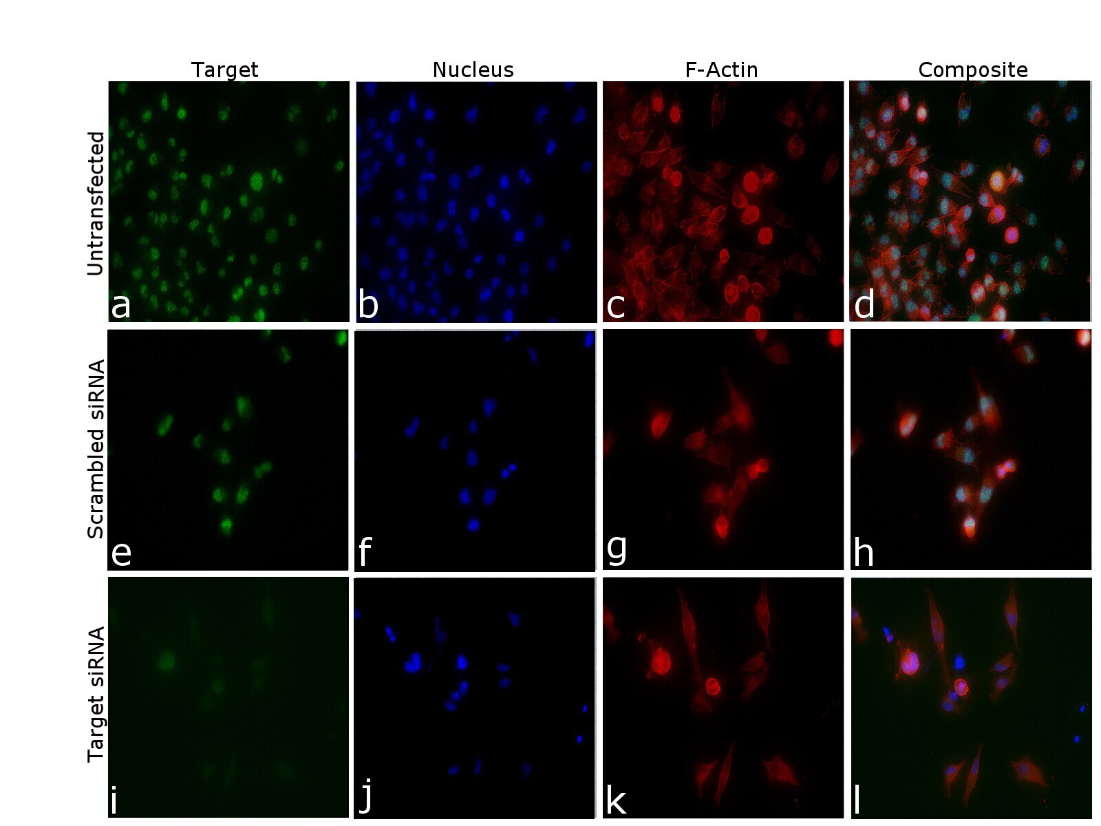
- Knockdown of JMJD1B was achieved by transfecting IMR-32 cells with JMJD1B specific siRNA (Silencer® select Product # s28657, s224243). Immunofluorescence analysis was performed on untransfected IMR-32 cells (panel a,d), transfected with non-specific scrambled siRNA (panels b,e) and transfected with JMJD1B specific siRNA (panel c,f). Cells were fixed, permeabilized, and labelled with JMJD1B Monoclonal Antibody (S.393.0) (Product # MA5-14792, 1:200 dilution) followed by Donkey anti-Rabbit IgG (H+L) Highly Cross-Adsorbed Secondary Antibody, Alexa Fluor Plus 488 (Product # A32790), (Donkey anti-Rabbit). Nuclei (blue) were stained using ProLong™ Diamond Antifade Mountant with DAPI (Product # P36962), and Rhodamine Phalloidin (Product # R415, 1:300) was used for cytoskeletal F-actin (Red) staining. Significant reduction of specific signal was observed upon siRNA mediated knockdown (panel i, k) confirming specificity of the antibody to JMJD1B (Green). The images were captured at 40X magnification in EVOS™ M7000 Imaging System (Product # AMF7000) and externally deconvoluted (D.Sage et al. / Methods 115 (2017) 28-41).

- Submitted by
- Invitrogen Antibodies (provider)
- Main image

- Experimental details
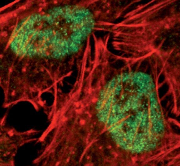
- Immunofluorescent analysis of JMJD1B in HeLa cells using a JMJD1B monoclonal antibody (Product # MA5-14792) (green). Actin filaments are labeled with a fluorescent red phalloidin.
- Submitted by
- Invitrogen Antibodies (provider)
- Main image

- Experimental details
- Immunofluorescent analysis of JMJD1B in HeLa cells using a JMJD1B monoclonal antibody (Product # MA5-14792) (green). Actin filaments are labeled with a fluorescent red phalloidin.

- Submitted by
- Invitrogen Antibodies (provider)
- Main image

- Experimental details
- Immunofluorescence analysis of JMJD1B was performed using 70% confluent log phase HCT 116 cells. The cells were fixed with 4% paraformaldehyde for 10 minutes, permeabilized with 0.1% Triton™ X-100 for 15 minutes, and blocked with 2% BSA for 1 hour at room temperature. The cells were labeled with JMJD1B Monoclonal Antibody (S.393.0) (Product # MA5-14792) at 1:100 dilution in 0.1% BSA, incubated at 4 degree celsius overnight and then labeled with Goat anti-Rabbit IgG (Heavy Chain) Superclonal™ Recombinant Secondary Antibody, Alexa Fluor® 488 conjugate (Product # A27034), (1:2000 dilution), for 45 minutes at room temperature (Panel a: Green). Nuclei (Panel b:Blue) were stained with ProLong™ Diamond Antifade Mountant with DAPI (Product # P36962). F-actin (Panel c: Red) was stained with Rhodamine Phalloidin (Product # R415, 1:300). Panel d represents the merged image showing nuclear localization. Panel e represents control cells with no primary antibody to assess background. The images were captured at 60X magnification.
- Submitted by
- Invitrogen Antibodies (provider)
- Main image

- Experimental details
- Knockdown of JMJD1B was achieved by transfecting IMR-32 cells with JMJD1B specific siRNA (Silencer® select Product # s28657, s224243). Immunofluorescence analysis was performed on untransfected IMR-32 cells (panel a,d), transfected with non-specific scrambled siRNA (panels b,e) and transfected with JMJD1B specific siRNA (panel c,f). Cells were fixed, permeabilized, and labelled with JMJD1B Monoclonal Antibody (S.393.0) (Product # MA5-14792, 1:200 dilution) followed by Donkey anti-Rabbit IgG (H+L) Highly Cross-Adsorbed Secondary Antibody, Alexa Fluor Plus 488 (Product # A32790), (Donkey anti-Rabbit). Nuclei (blue) were stained using ProLong™ Diamond Antifade Mountant with DAPI (Product # P36962), and Rhodamine Phalloidin (Product # R415, 1:300) was used for cytoskeletal F-actin (Red) staining. Significant reduction of specific signal was observed upon siRNA mediated knockdown (panel i, k) confirming specificity of the antibody to JMJD1B (Green). The images were captured at 40X magnification in EVOS™ M7000 Imaging System (Product # AMF7000) and externally deconvoluted (D.Sage et al. / Methods 115 (2017) 28-41).
Supportive validation
- Submitted by
- Invitrogen Antibodies (provider)
- Main image

- Experimental details
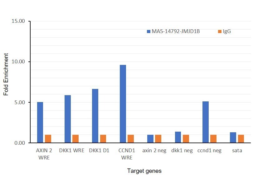
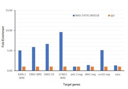
- Chromatin Immunoprecipitation (ChIP) was performed using JMJD1B Monoclonal Antibody (S.393.0) (Product # MA5-14792, 10 ul) on sheared chromatin from a million HCT 116 cells using the MAGnify ChIP System (Product # 49-2024). Normal Rabbit IgG was used as a negative IP control. The purified DNA was analyzed by qPCR with PCR primer pairs over Axin 2 WRE region, DKK1 WRE region, DKK1 D1 region, CCND1 WRE genes (active), and Axin 2 Neg, DKK1 neg,CCND1 neg and SAT2 satellite repeats (inactive). Antibody specificity was demonstrated by detection of enrichment of the target protein at specific gene loci and no enrichment seen in negative loci except for CCND1 where there is low enrichment in comparison to CCND1 WRE region. Data is presented as fold enrichment of the antibody signal versus the Rabbit Isotype using the comparative CT method.

- Submitted by
- Invitrogen Antibodies (provider)
- Main image
- Experimental details
- Chromatin Immunoprecipitation (ChIP) was performed using JMJD1B Monoclonal Antibody (S.393.0) (Product # MA5-14792, 10 ul) on sheared chromatin from a million HCT 116 cells using the MAGnify ChIP System (Product # 49-2024). Normal Rabbit IgG was used as a negative IP control. The purified DNA was analyzed by qPCR with PCR primer pairs over Axin 2 WRE region, DKK1 WRE region, DKK1 D1 region, CCND1 WRE genes (active), and Axin 2 Neg, DKK1 neg,CCND1 neg and SAT2 satellite repeats (inactive). Antibody specificity was demonstrated by detection of enrichment of the target protein at specific gene loci and no enrichment seen in negative loci except for CCND1 where there is low enrichment in comparison to CCND1 WRE region. Data is presented as fold enrichment of the antibody signal versus the Rabbit Isotype using the comparative CT method.