Explore
 Validate
 Learn
LearnAP09259PU-N
antibody from Acris Antibodies GmbH
Targeting: EGR1
AT225, G0S30, KROX-24, NGFI-A, TIS8, ZIF-268, ZNF225
 Western blot
 ELISA
ELISAAntibody data
- Antibody Data
- Antigen structure
- References [0]
- Comments [0]
- Validations
- Western blot [1]
- Immunohistochemistry [1]
Submit
Validation data
Reference
Comment
Report error
- Product number
- AP09259PU-N - Provider product page

- Provider
- Acris Antibodies GmbH
- Proper citation
- Acris Antibodies GmbH Cat#AP09259PU-N, RRID:AB_2035450
- Product name
- anti EGR1 (94-108)
- Antibody type
- Polyclonal
- Antigen
- Synthetic peptide corresponding to amino acids 94-108 of Human EGR-1
- Reactivity
- Human, Mouse, Rat
- Host
- Rabbit
- Isotype
- IgG
- Vial size
- 0.1 mg
- Concentration
- 1.46 mg/ml (by UV absorbance at 280 nm)
No comments: Submit comment
Supportive validation
- Submitted by
- Acris Antibodies GmbH (provider)
- Main image
- Experimental details
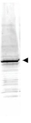
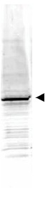
- Western blot using Anti-EGR-1 antibody shows detection of a predominant band at ~58 kDa corresponding to EGR-1 present in mouse embryonic fibroblast whole cell lysate (arrowhead). Approximately 35 μg of lysate was separated by 4-20% SDS-PAGE and transferred onto nitrocellulose. After blocking the membrane was probed with the primary antibody diluted to 1:1,500. Reaction occurred 2h at room temperature followed by washes and reaction with a 1:10,000 dilution of IRDye(TM)800 conjugated Gta-Rabbit IgG [H&L] for 45 min at room temperature. IRDye(TM)800 fluorescence image was captured using the Odyssey® Infrared Imaging System developed by LI-COR. IRDye is a trademark of LI-COR, Inc. Other detection systems will yield similar results.
Supportive validation
- Submitted by
- Acris Antibodies GmbH (provider)
- Main image

- Experimental details
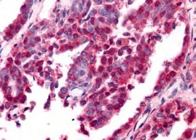
- Immunohistochemistry. Anti-EGR-1 antibody was used at a 10 μg/ml to detect nuclear and cytoplasmic signal with low background staining in a variety of tissues including multi-human, multi-brain and multi-cancer slides. Within the multi-tumor block, the antibody showed variable levels of nuclear and cytoplasmic staining between individual tumors, with some tumors showing moderate staining. This image shows EGR-1 staining of human ovarian carcinoma. Tissue was formalin-fixed and paraffin embedded. Personal Communication, Tina Roush, LifeSpanBiosciences, Seattle, WA.