Explore
 Validate
 Learn
Learn Western blot
Antibody data
- Antibody Data
- Antigen structure
- References [0]
- Comments [0]
- Validations
- Western blot [1]
- Immunohistochemistry [1]
Submit
Validation data
Reference
Comment
Report error
- Product number
- TA319423 - Provider product page

- Provider
- OriGene
- Product name
- Rabbit polyclonal anti-EGR-1 antibody
- Antibody type
- Polyclonal
- Description
- Rabbit polyclonal anti-EGR-1 antibody
- Host
- Rabbit
- Conjugate
- Unconjugated
- Epitope
- EGR1
- Isotype
- IgG
- Antibody clone number
- NULL
- Vial size
- 100 µg
- Concentration
- 0.93 mg/mL
No comments: Submit comment
Supportive validation
- Submitted by
- OriGene (provider)
- Main image

- Experimental details
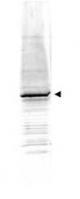
- WB using Anti-EGR-1 antibody shows detection of a predominant band at ~58 kDa corresponding to EGR-1 present in mouse embryonic fibroblast whole cell lysate (arrowhead). Primary antibody was used at a 1,500 dilution. Reaction occurred 2h at room temperature followed by washes and reaction with a 1:10,000 dilution of IRDye?800 conjugated Gt-a-Rabbit IgG [H&L] MX for 45 min at room temperature.
- Validation comment
- WB
Supportive validation
- Submitted by
- OriGene (provider)
- Main image

- Experimental details
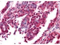
- Anti-EGR-1 antibody was used at a 10 ug/ml to detect nuclear and cytoplasmic signal with low background staining in a variety of tissues including multi-human, multi-brain and multi-cancer slides. Within the multi-tumor block, the antibody showed variable levels of nuclear and cytoplasmic staining between individual tumors, with some tumors showing moderate staining. This image shows EGR-1 staining of human ovarian carcinoma. Tissue was formalin-fixed and paraffin embedded.
- Validation comment
- IHC