Explore
 Validate
 Learn
Learn Immunocytochemistry
Antibody data
- Antibody Data
- Antigen structure
- References [1]
- Comments [0]
- Validations
- Immunocytochemistry [1]
- Immunohistochemistry [1]
Submit
Validation data
Reference
Comment
Report error
- Product number
- HPA055223 - Provider product page

- Provider
- Atlas Antibodies
- Proper citation
- Atlas Antibodies Cat#HPA055223, RRID:AB_2682745
- Product name
- Anti-TCFL5
- Antibody type
- Polyclonal
- Description
- Polyclonal Antibody against Human TCFL5, Gene description: transcription factor-like 5 (basic helix-loop-helix), Alternative Gene Names: bHLHe82, CHA, E2BP-1, Figlb, Validated applications: ICC, IHC, Uniprot ID: Q9UL49, Storage: Store at +4°C for short term storage. Long time storage is recommended at -20°C.
- Reactivity
- Human
- Host
- Rabbit
- Conjugate
- Unconjugated
- Isotype
- IgG
- Vial size
- 100 µl
- Concentration
- 0.1 mg/ml
- Storage
- Store at +4°C for short term storage. Long time storage is recommended at -20°C.
- Handling
- The antibody solution should be gently mixed before use.
Submitted references A-MYB/TCFL5 regulatory architecture ensures the production of pachytene piRNAs in placental mammals
Yu T, Biasini A, Cecchini K, Säflund M, Mou H, Arif A, Eghbali A, de Rooij D, Weng Z, Zamore P, Özata D
RNA 2023;29(1):30-43
RNA 2023;29(1):30-43
No comments: Submit comment
Supportive validation
- Submitted by
- Atlas Antibodies (provider)
- Main image
- Experimental details
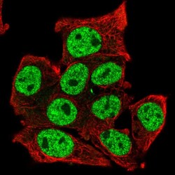
- Immunofluorescent staining of human cell line Hep G2 shows localization to nucleoplasm.
- Sample type
- Human

Supportive validation
- Submitted by
- Atlas Antibodies (provider)
- Enhanced method
- Orthogonal validation
- Main image
- Experimental details
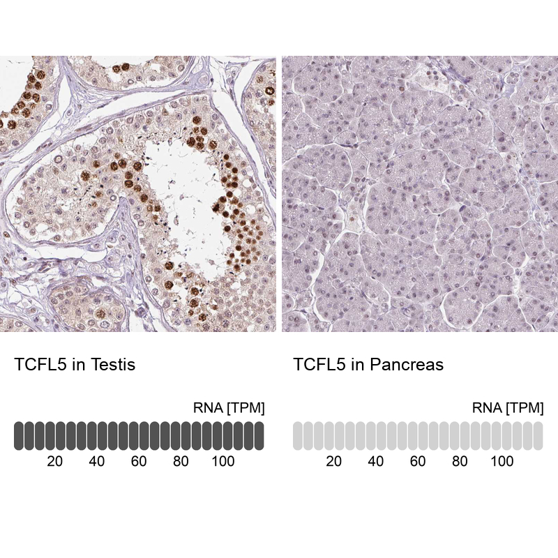
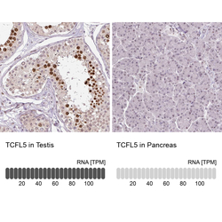
- Immunohistochemistry analysis in human testis and pancreas tissues using HPA055223 antibody. Corresponding TCFL5 RNA-seq data are presented for the same tissues.
- Sample type
- Human
- Protocol
- Protocol