Explore
 Validate
 Learn
Learn Immunohistochemistry
Antibody data
- Antibody Data
- Antigen structure
- References [3]
- Comments [0]
- Validations
- Immunohistochemistry [3]
Submit
Validation data
Reference
Comment
Report error
- Product number
- 14-9761-80 - Provider product page

- Provider
- Invitrogen Antibodies
- Product name
- CD11c Monoclonal Antibody (118/A5), eBioscience™
- Antibody type
- Monoclonal
- Antigen
- Other
- Description
- Description: The monoclonal antibody 118/A5 recognizes human CD11c, a 150 kD type I transmembrane protein which is also known as integrin alpha X. Integrins are heterodimeric integral membrane proteins made up of an alpha and a beta chain and are involved in cell adhesion. The CD11/CD18 adhesion molecules consist of three heterodimers sharing a common beta subunit (CD18) with a distinct alpha subunit (CD11a, CD11b, CD11c). The CD11c/CD18 complex is expressed on dendritic cells (highly expressed on type 1 myeloid DCs), monocytes, macrophages, granulocytes, NK cells, and subsets of T and B cells. CD11c is also expressed in hairy cell leukemias, acute nonlymphocytic leukemias, and some B-cell chronic lymphocytic leukemias. CD11c plays a role in adhesion and CTL killing through its interactions with CD54, iC3b and fibrinogen. Applications Reported: This 118/A5 antibody has been reported for use in immunohistochemical staining of frozen tissue sections, immunohistochemical staining of formalin-fixed paraffin embedded tissue sections, and microscopy. Applications Tested: This 118/A5 antibody has been tested by immunohistochemistry of formalin-fixed paraffin embedded human tissue using low or high pH antigen retrieval and can be used at less than or equal to 5 µg/mL. It is recommended that the antibody be carefully titrated for optimal performance in the assay of interest. Purity: Greater than 90%, as determined by SDS-PAGE. Aggregation: Less than 10%, as determined by HPLC. Filtration: 0.2 µm post-manufacturing filtered.
- Reactivity
- Human
- Host
- Mouse
- Isotype
- IgG
- Antibody clone number
- 118/A5
- Vial size
- 25 μg
- Concentration
- 0.5 mg/mL
- Storage
- 4°C
Submitted references Comparative analysis of integrin expression on monocyte-derived macrophages and monocyte-derived dendritic cells.
The leukocyte integrin p150,95 (CD11c/CD18) as a receptor for iC3b. Activation by a heterologous beta subunit and localization of a ligand recognition site to the I domain.
The monoclonal antibodies alpha S-HCL 1 (alpha Leu-14) and alpha S-HCL 3 (alpha Leu-M5) allow the diagnosis of hairy cell leukemia.
Ammon C, Meyer SP, Schwarzfischer L, Krause SW, Andreesen R, Kreutz M
Immunology 2000 Jul;100(3):364-9
Immunology 2000 Jul;100(3):364-9
The leukocyte integrin p150,95 (CD11c/CD18) as a receptor for iC3b. Activation by a heterologous beta subunit and localization of a ligand recognition site to the I domain.
Bilsland CA, Diamond MS, Springer TA
Journal of immunology (Baltimore, Md. : 1950) 1994 May 1;152(9):4582-9
Journal of immunology (Baltimore, Md. : 1950) 1994 May 1;152(9):4582-9
The monoclonal antibodies alpha S-HCL 1 (alpha Leu-14) and alpha S-HCL 3 (alpha Leu-M5) allow the diagnosis of hairy cell leukemia.
Schwarting R, Stein H, Wang CY
Blood 1985 Apr;65(4):974-83
Blood 1985 Apr;65(4):974-83
No comments: Submit comment
Supportive validation
- Submitted by
- Invitrogen Antibodies (provider)
- Main image
- Experimental details
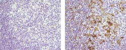
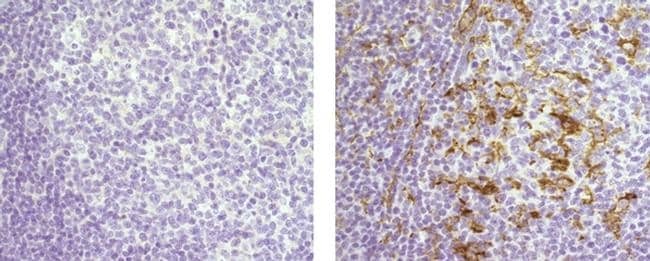
- Immunohistochemistry of formalin-fixed paraffin embedded human tonsil using 5 µg/mL Mouse IgG2b K Isotype Control Purified (left) or 5 µg/mL Anti-Human CD11c Purified (right) followed by Anti-Mouse IgG Biotin and Streptavidin HRP.Nuclei are counterstained with hematoxylin.
- Submitted by
- Invitrogen Antibodies (provider)
- Main image
- Experimental details
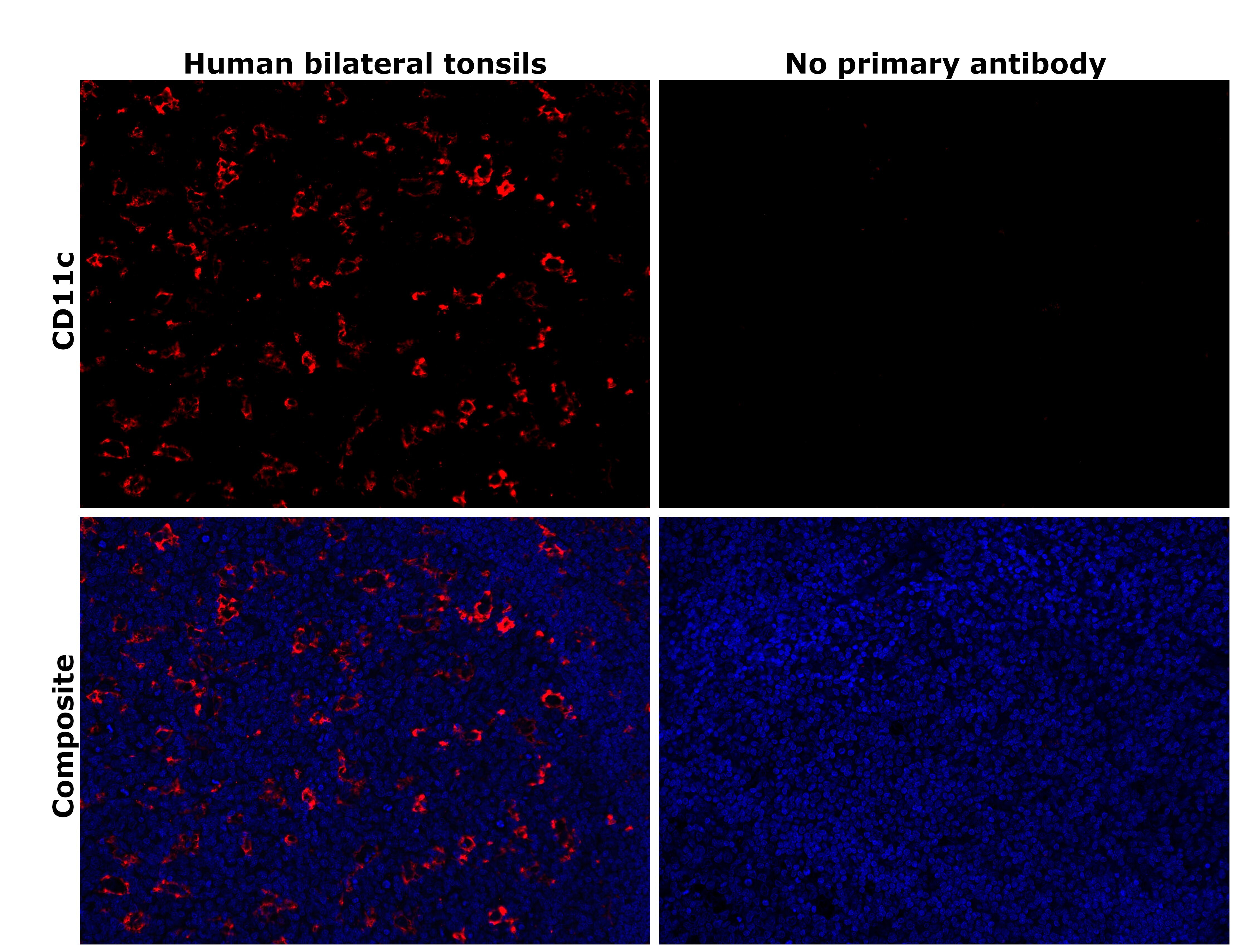
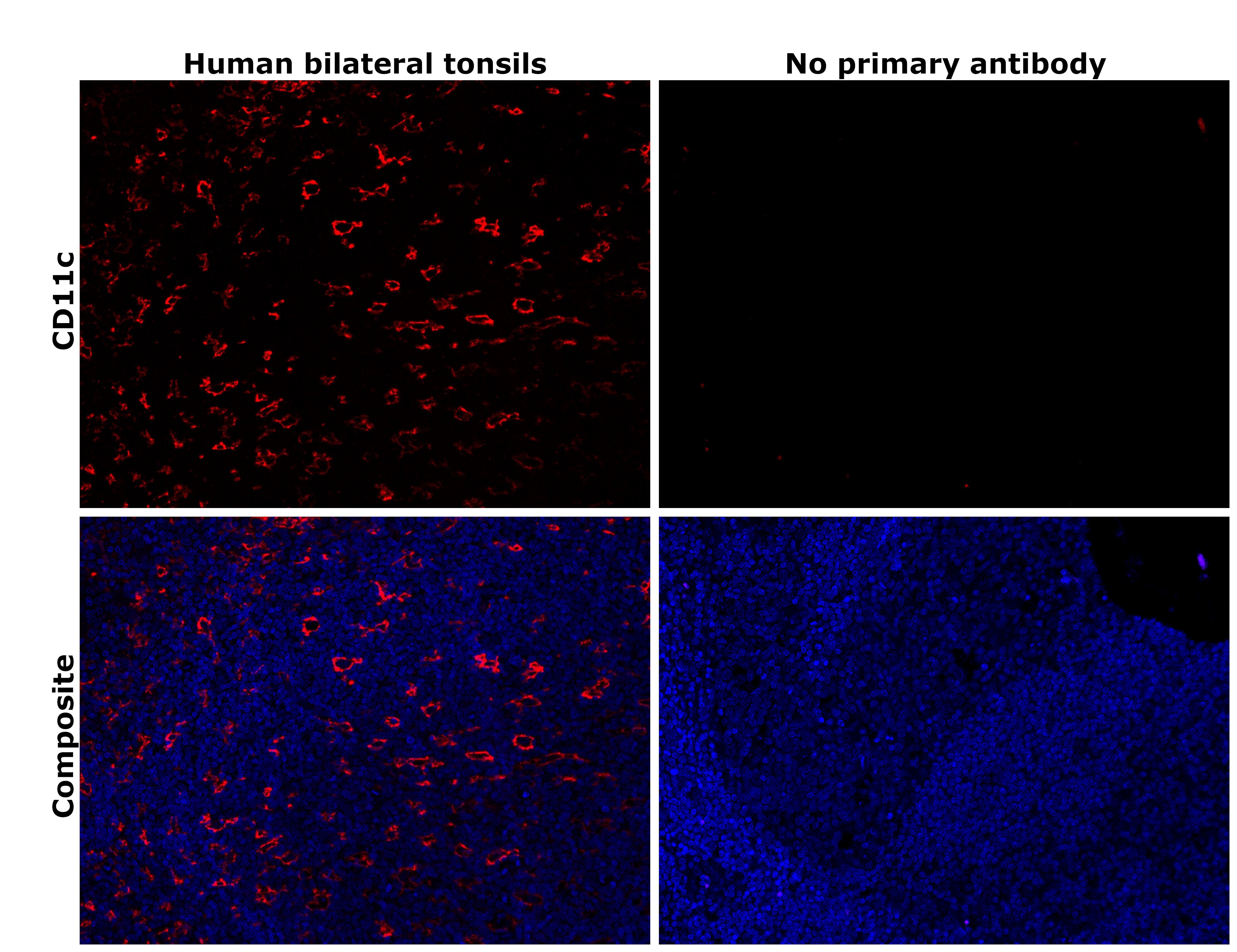
- Immunohistochemical analysis of CD11c was performed using formalin-fixed paraffin-embedded human bilateral tonsils tissue sections. To expose the target protein, heat-induced epitope retrieval was performed on de-paraffinized sections using eBioscience™ IHC Antigen Retrieval Solution - High pH (10X) (Product # 00-4956-58) diluted to 1X solution in water in a decloaking chamber at 110 degree Celsius for 15 minutes. Following antigen retrieval, the sections were blocked with 3% H2O2 for 1 hour at room temperature followed by 2% normal goat serum in 1X PBS for 45 minutes at room temperature and then probed with or without CD11c Monoclonal Antibody (118/A5), eBioscience™ (Product # 14-9761-82) at 1 µg/mL in 0.1% normal goat serum overnight at 4 degree Celsius in a humidified chamber. Detection was performed using Alexa Fluor™ 555 Tyramide SuperBoost™ Kit, goat anti-mouse IgG (Product # B40913). Nuclei were stained with DAPI (Product # D1306) and the sections were mounted using ProLong™ Glass Antifade Mountant (Product # P36984). The images were captured on EVOS™ M7000 Imaging System (Product # AMF7000) at 20X magnification and externally deconvoluted.
- Submitted by
- Invitrogen Antibodies (provider)
- Main image
- Experimental details
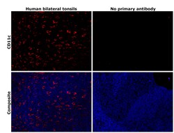
- Immunohistochemical analysis of CD11c was performed using formalin-fixed paraffin-embedded human bilateral tonsils tissue sections. To expose the target protein, heat-induced epitope retrieval was performed on de-paraffinized sections using eBioscience™ IHC Antigen Retrieval Solution - Low pH (10X) (Product # 00-4955-58) diluted to 1X solution in water in a decloaking chamber at 110 degree Celsius for 15 minutes. Following antigen retrieval, the sections were blocked with 3% H2O2 for 1 hour at room temperature followed by 2% normal goat serum in 1X PBS for 45 minutes at room temperature and then probed with or without CD11c Monoclonal Antibody (118/A5), eBioscience™ (Product # 14-9761-82) at 1 µg/mL in 0.1% normal goat serum overnight at 4 degree Celsius in a humidified chamber. Detection was performed using Alexa Fluor™ 555 Tyramide SuperBoost™ Kit, goat anti-mouse IgG (Product # B40913). Nuclei were stained with DAPI (Product # D1306) and the sections were mounted using ProLong™ Glass Antifade Mountant (Product # P36984). The images were captured on EVOS™ M7000 Imaging System (Product # AMF7000) at 20X magnification and externally deconvoluted.