 Explore
 Validate
 Learn
Learn Immunohistochemistry
Antibody data
- Antibody Data
- Antigen structure
- References [0]
- Comments [0]
- Validations
- Immunohistochemistry [1]
- Flow cytometry [1]
Submit
Validation data
Reference
Comment
Report error
- Product number
- 10-7566-25 - Provider product page

- Provider
- ABEOMICS Inc.
- Product name
- Anti-CD11c Antibody
- Antibody type
- Monoclonal
- Description
- CD11c/CD18 ( p150/95, or complement receptor 4, CR4) is a monocyte/macrophage-enriched integrin that has been reported to bind to a variety of ligands. These include cell surface proteins (LPS, ICAM-1, ICAM-2, ICAM-4 and VCAM-1), extracellular matrix proteins (collagen I), and soluble ligands (iC3b, heparin and fibrinogen). It is expressed in macrophages, monocytes, granulocytes, subsets of T and B cells, and dendritic cells. CD11c functions as a cell surface receptor for numerous soluble factors and proteins. The interaction mediates leukocyte interactions with other cell types and is a signal transducing receptor. It is found primarily on myeloid cells, where its expression is regulated both during differentiation and during monocyte maturation into tissue macrophages.
- Reactivity
- Human
- Host
- Mouse
- Conjugate
- Unconjugated
- Antigen sequence
A partial length recombinant CD11c
protein (amino acids 620-835) was u
sed as the immunogen for this antib
ody.- Isotype
- IgG
- Antibody clone number
- ABM4C58
- Vial size
- 100 µg
- Concentration
- 0.5 mg/ml
- Storage
- Store the antibody at 4°C, stable for 6 months. For long-term storage, store at -20°C. Avoid repeat freez thawing
No comments: Submit comment
Supportive validation
- Submitted by
- ABEOMICS Inc. (provider)
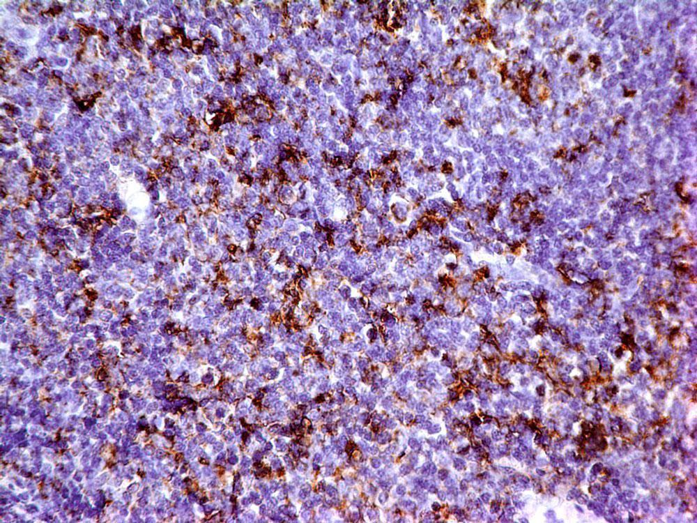
- Main image
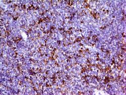
- Experimental details
- Immunohistochemical analysis of CD11c in human Tonsil tissue using CD11c antibody (Clone: ABM4C58) at 5 µg/ml.
- Protocol
- Protocol
Supportive validation
- Submitted by
- ABEOMICS Inc. (provider)
- Main image
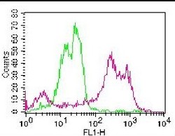
- Experimental details
- Cell surface flow analysis of hCD11C on human PBMCs using 1 µg/ 10^6 cells. Green represents isotype control (ABEOMICS); red represents anti-hCD11C antibody (10-7566). Goat anti-mouse FITC conjugated secondary antibody (ABEOMICS) was used. (Cells were incubated with primary antibody for 30 min. then washed twice with FLOW Staining buffer (ABEOMICS) by centrifuging at 1000 rpm for 5 min, followed by 30 min incubation with conjugated secondary antibody. Data acquisition was done after washing twice with FLOW staining buffer).
- Protocol
- Protocol
