Explore
 Validate
 Learn
Learn Western blot
Antibody data
- Antibody Data
- Antigen structure
- References [1]
- Comments [0]
- Validations
- Western blot [3]
- Immunocytochemistry [2]
- Immunohistochemistry [1]
- Flow cytometry [1]
Submit
Validation data
Reference
Comment
Report error
- Product number
- TA500739 - Provider product page

- Provider
- OriGene
- Proper citation
- OriGene Cat#TA500739, RRID:AB_11141358
- Product name
- Anti-IDH3A mouse monoclonal antibody, clone OTI2F11 (formerly 2F11)
- Antibody type
- Monoclonal
- Description
- Anti-IDH3A mouse monoclonal antibody, clone OTI2F11 (formerly 2F11)
- Reactivity
- Canine
- Host
- Mouse
- Conjugate
- Unconjugated
- Epitope
- IDH3A
- Isotype
- IgG
- Antibody clone number
- OTI2F11
- Vial size
- 100 µl
- Concentration
- 1.00mg/ml
Submitted references Metabolic reprogramming of cancer-associated fibroblasts by IDH3α downregulation.
Zhang D, Wang Y, Shi Z, Liu J, Sun P, Hou X, Zhang J, Zhao S, Zhou BP, Mi J
Cell reports 2015 Mar 3;10(8):1335-48
Cell reports 2015 Mar 3;10(8):1335-48
No comments: Submit comment
Supportive validation
- Submitted by
- OriGene (provider)
- Main image
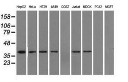
- Experimental details
- Western blot analysis of extracts (35ug) from 9 different cell lines by using anti-IDH3A monoclonal antibody.
- Validation comment
- WB

- Submitted by
- OriGene (provider)
- Main image
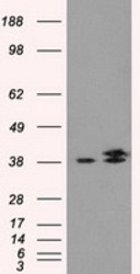
- Experimental details
- HEK293T cells were transfected with the pCMV6-ENTRY control (Left lane) or pCMV6-ENTRY IDH3A (RC200313, Right lane) cDNA for 48 hrs and lysed. Equivalent amounts of cell lysates (5 ug per lane) were separated by SDS-PAGE and immunoblotted with anti-IDH3A.
- Validation comment
- WB
- Submitted by
- OriGene (provider)
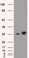
- Main image
- Experimental details
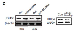
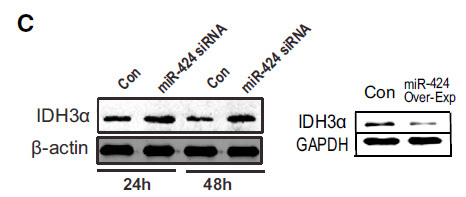
- Figure from citation: Western blot analysis of IDH3A protein level by using anti-IDH3A antibody in fibroblasts with miR-424 knockdown or overexpression,and in TGF-b1-treated fibroblasts with or without miR-424 depletion.
- Validation comment
- WB
Supportive validation
- Submitted by
- OriGene (provider)
- Main image
- Experimental details
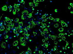
- Anti-IDH3A mouse monoclonal antibody (TA500739) immunofluorescent staining of COS7 cells transiently transfected by pCMV6-ENTRY IDH3A(RC200313).
- Validation comment
- IF
- Submitted by
- OriGene (provider)
- Main image
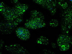
- Experimental details
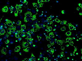
- Immunofluorescent staining of HepG2 cells using anti-IDH3A mouse monoclonal antibody (TA500739).
- Validation comment
- IF

Supportive validation
- Submitted by
- OriGene (provider)
- Main image

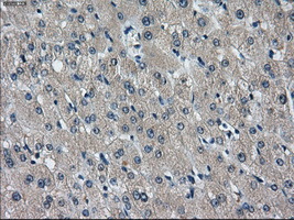
- Experimental details
- Immunohistochemical staining of paraffin-embedded liver tissue within the normal limits using anti-IDH3Amouse monoclonal antibody. (Heat-induced epitope retrieval by 10mM citric buffer, pH6.0, 100C for 10min, TA500739, Dilution 1:50)
- Validation comment
- IHC
Supportive validation
- Submitted by
- OriGene (provider)
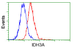
- Main image
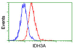
- Experimental details
- Flow cytometric analysis of Jurkat cells, using anti-IDH3A antibody(TA500739),(Red) compared to a nonspecific negative control antibody(TA50011)(Blue).
- Validation comment
- FC