Explore
 Validate
 Learn
Learn Western blot
 Immunocytochemistry
Antibody data
- Antibody Data
- Antigen structure
- References [9]
- Comments [0]
- Validations
- Western blot [1]
- Immunocytochemistry [1]
- Immunohistochemistry [1]
Submit
Validation data
Reference
Comment
Report error
- Product number
- HPA000992 - Provider product page

- Provider
- Atlas Antibodies
- Proper citation
- Atlas Antibodies Cat#HPA000992, RRID:AB_1079009
- Product name
- Anti-GOLGA5
- Antibody type
- Polyclonal
- Description
- Polyclonal Antibody against Human GOLGA5, Gene description: golgin A5, Alternative Gene Names: golgin-84, GOLIM5, ret-II, rfg5, Validated applications: ICC, IHC, WB, Uniprot ID: Q8TBA6, Storage: Store at +4°C for short term storage. Long time storage is recommended at -20°C.
- Reactivity
- Human
- Host
- Rabbit
- Conjugate
- Unconjugated
- Isotype
- IgG
- Vial size
- 100 µl
- Concentration
- 0.1 mg/ml
- Storage
- Store at +4°C for short term storage. Long time storage is recommended at -20°C.
- Handling
- The antibody solution should be gently mixed before use.
Submitted references Dynamic movement of the Golgi unit and its glycosylation enzyme zones
DJ-1 is an essential downstream mediator in PINK1/parkin-dependent mitophagy
Direct homophilic interaction of LAMP2A with the two-domain architecture revealed by site-directed photo-crosslinks and steric hindrances in mammalian cells
In vivo identification of GTPase interactors by mitochondrial relocalization and proximity biotinylation
The golgin coiled-coil proteins capture different types of transport carriers via distinct N-terminal motifs
The specificity of vesicle traffic to the Golgi is encoded in the golgin coiled-coil proteins
Systematic validation of antibody binding and protein subcellular localization using siRNA and confocal microscopy
Multiple domains in the Crumbs Homolog 2a (Crb2a) protein are required for regulating rod photoreceptor size
Systematically generated antibodies against human gene products: High throughput screening on sections from the rat nervous system
Harada A, Kunii M, Kurokawa K, Sumi T, Kanda S, Zhang Y, Nadanaka S, Hirosawa K, Tokunaga K, Tojima T, Taniguchi M, Moriwaki K, Yoshimura S, Yamamoto-Hino M, Goto S, Katagiri T, Kume S, Hayashi-Nishino M, Nakano M, Miyoshi E, Suzuki K, Kitagawa H, Nakano A
Nature Communications 2024;15(1)
Nature Communications 2024;15(1)
DJ-1 is an essential downstream mediator in PINK1/parkin-dependent mitophagy
Imberechts D, Kinnart I, Wauters F, Terbeek J, Manders L, Wierda K, Eggermont K, Madeiro R, Sue C, Verfaillie C, Vandenberghe W
Brain 2022;145(12):4368-4384
Brain 2022;145(12):4368-4384
Direct homophilic interaction of LAMP2A with the two-domain architecture revealed by site-directed photo-crosslinks and steric hindrances in mammalian cells
Terasawa K, Kato Y, Ikami Y, Sakamoto K, Ohtake K, Kusano S, Tomabechi Y, Kukimoto-Niino M, Shirouzu M, Guan J, Kobayashi T, Iwata T, Watabe T, Yokoyama S, Hara-Yokoyama M
Autophagy 2021;17(12):4286-4304
Autophagy 2021;17(12):4286-4304
In vivo identification of GTPase interactors by mitochondrial relocalization and proximity biotinylation
Gillingham A, Bertram J, Begum F, Munro S
eLife 2019;8
eLife 2019;8
The golgin coiled-coil proteins capture different types of transport carriers via distinct N-terminal motifs
Wong M, Gillingham A, Munro S
BMC Biology 2017;15(1)
BMC Biology 2017;15(1)
The specificity of vesicle traffic to the Golgi is encoded in the golgin coiled-coil proteins
Wong M, Munro S
Science 2014;346(6209)
Science 2014;346(6209)
Systematic validation of antibody binding and protein subcellular localization using siRNA and confocal microscopy
Stadler C, Hjelmare M, Neumann B, Jonasson K, Pepperkok R, Uhlén M, Lundberg E
Journal of Proteomics 2012;75(7):2236-2251
Journal of Proteomics 2012;75(7):2236-2251
Multiple domains in the Crumbs Homolog 2a (Crb2a) protein are required for regulating rod photoreceptor size
Hsu Y, Jensen A
BMC Cell Biology 2010;11(1)
BMC Cell Biology 2010;11(1)
Systematically generated antibodies against human gene products: High throughput screening on sections from the rat nervous system
Mulder J, Wernérus H, Shi T, Pontén F, Hober S, Uhlén M, Hökfelt T
Neuroscience 2007;146(4):1689-1703
Neuroscience 2007;146(4):1689-1703
No comments: Submit comment
Enhanced validation
- Submitted by
- Atlas Antibodies (provider)
- Enhanced method
- Genetic validation
- Main image
- Experimental details
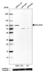
- Western blot analysis in U-87MG ATCC cells transfected with control siRNA, target specific siRNA probe #1 and #2, using Anti-GOLGA5 antibody. Remaining relative intensity is presented. Loading control: Anti-GAPDH.
- Sample type
- Human
- Protocol
- Protocol

Supportive validation
- Submitted by
- Atlas Antibodies (provider)
- Main image

- Experimental details
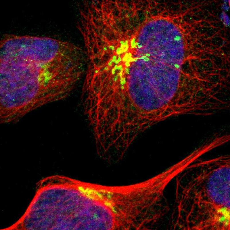
- Immunofluorescent staining of human cell line U-2 OS shows localization to the Golgi apparatus.
- Sample type
- Human
Supportive validation
- Submitted by
- Atlas Antibodies (provider)
- Enhanced method
- Orthogonal validation
- Main image

- Experimental details
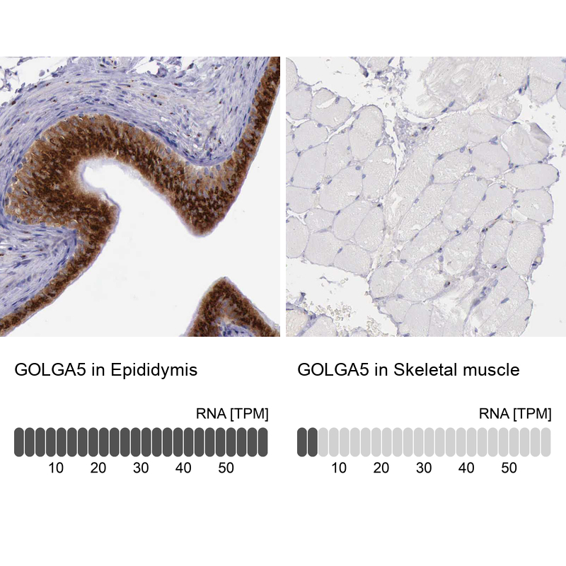
- Immunohistochemistry analysis in human epididymis and skeletal muscle tissues using HPA000992 antibody. Corresponding GOLGA5 RNA-seq data are presented for the same tissues.
- Sample type
- Human
- Protocol
- Protocol