Antibody data
- Antibody Data
- Antigen structure
- References [0]
- Comments [0]
- Validations
- Western blot [2]
- Immunohistochemistry [10]
Submit
Validation data
Reference
Comment
Report error
- Product number
- HPA008877 - Provider product page
- Provider
- Atlas Antibodies
- Proper citation
- Atlas Antibodies Cat#HPA008877, RRID:AB_1846256
- Product name
- Anti-ITGB2
- Antibody type
- Polyclonal
- Reactivity
- Human
- Host
- Rabbit
- Conjugate
- Unconjugated
- Antigen sequence
CTKFKVSSCRECIESGPGCTWCQKLNFTGPGDPDS
IRCDTRPQLLMRGCAADDIMDPTSLAETQEDHNGG
QKQLSPQKVTLYLRPGQAAAFNVTFRRAKGYPIDL
YYLMDLSYSML- Isotype
- IgG
- Vial size
- 100 µl
- Storage
- Store at +4°C for short term storage. Long time storage is recommended at -20°C.
No comments: Submit comment
Supportive validation
- Submitted by
- Atlas Antibodies (provider)
- Enhanced method
- Independent antibody validation
- Main image
- Experimental details
- Western blot analysis using Anti-ITGB2 antibody HPA008877 (A) shows similar pattern to independent antibody HPA016894 (B).
Supportive validation
- Submitted by
- Atlas Antibodies (provider)
- Main image
- Experimental details
- Lane 1: Marker [kDa] 230, 130, 95, 72, 56, 36, 28, 17, 11Lane 2: Human cell line RT-4Lane 3: Human cell line U-251MG spLane 4: Human plasma (IgG/HSA depleted)Lane 5: Human liver tissueLane 6: Human tonsil tissue
- Sample type
- HUMAN
Enhanced validation
Enhanced validation
Supportive validation
- Submitted by
- Atlas Antibodies (provider)
- Enhanced method
- Orthogonal validation
- Main image
- Experimental details
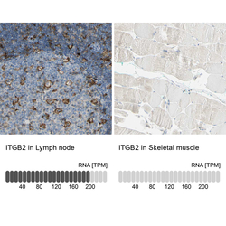
- Immunohistochemistry analysis in human lymph node and skeletal muscle tissues using HPA008877 antibody. Corresponding ITGB2 RNA-seq data are presented for the same tissues.
- Sample type
- HUMAN
Enhanced validation
- Submitted by
- Atlas Antibodies (provider)
- Enhanced method
- Independent antibody validation
- Main image
- Experimental details
- Immunohistochemical staining of human gastrointestinal, lymph node, skeletal muscle and tonsil using Anti-ITGB2 antibody HPA008877 (A) shows similar protein distribution across tissues to independent antibody HPA016894 (B).
Supportive validation
- Submitted by
- Atlas Antibodies (provider)
- Main image
- Experimental details
- Immunohistochemical staining of human spleen shows high expression.
- Sample type
- HUMAN
- Submitted by
- Atlas Antibodies (provider)
- Main image
- Experimental details
- Immunohistochemical staining of human skeletal muscle shows low expression as expected.
- Sample type
- HUMAN
- Submitted by
- Atlas Antibodies (provider)
- Main image
- Experimental details
- Immunohistochemical staining of human lymph node using Anti-ITGB2 antibody HPA008877.
- Sample type
- HUMAN
- Submitted by
- Atlas Antibodies (provider)
- Main image
- Experimental details
- Immunohistochemical staining of human bone marrow using Anti-ITGB2 antibody HPA008877.
- Sample type
- HUMAN
- Submitted by
- Atlas Antibodies (provider)
- Main image
- Experimental details
- Immunohistochemical staining of human lymph node shows strong membranous positivity in germinal center cells.
- Sample type
- HUMAN
- Submitted by
- Atlas Antibodies (provider)
- Main image
- Experimental details
- Immunohistochemical staining of human skeletal muscle shows no positivity in myocytes as expected.
- Sample type
- HUMAN
- Submitted by
- Atlas Antibodies (provider)
- Main image
- Experimental details
- Immunohistochemical staining of human colon shows strong membranous positivity in lymphoid cells.
- Sample type
- HUMAN
- Submitted by
- Atlas Antibodies (provider)
- Main image
- Experimental details
- Immunohistochemical staining of human tonsil shows strong membranous positivity in germinal center cells.
- Sample type
- HUMAN