Explore
 Validate
 Learn
Learn Western blot
 ELISA
ELISAAntibody data
- Antibody Data
- Antigen structure
- References [8]
- Comments [0]
- Validations
- Western blot [1]
- Immunocytochemistry [1]
- Immunohistochemistry [1]
Submit
Validation data
Reference
Comment
Report error
- Product number
- 10886-1-AP - Provider product page

- Provider
- Proteintech Group
- Proper citation
- Proteintech Cat#10886-1-AP, RRID:AB_2182999
- Product name
- Syndecan-3 antibody
- Antibody type
- Polyclonal
- Description
- Syndecan-3 antibody (Cat. #10886-1-AP) is a rabbit polyclonal antibody that shows reactivity with human, mouse, rat and has been validated for the following applications: IF, IHC, IP, WB,ELISA.
- Reactivity
- Human, Mouse, Rat
- Host
- Rabbit
- Conjugate
- Unconjugated
- Isotype
- IgG
- Vial size
- 20ul, 150ul
Submitted references Long-Term High-Fat Diet Affected Bone Marrow Microenvironment During Aging at Single-Cell Resolution.
Two novel predictive biomarkers for osteosarcoma and glycolysis pathways: A profiling study on HS2ST1 and SDC3.
GDNF Increases Inhibitory Synaptic Drive on Principal Neurons in the Hippocampus via Activation of the Ret Pathway.
Metabolic and chemical regulation of tRNA modification associated with taurine deficiency and human disease.
Electrophoresis of cell membrane heparan sulfate regulates galvanotaxis in glial cells.
Prognostic significance of the expression of GFRα1, GFRα3 and syndecan-3, proteins binding ARTEMIN, in mammary carcinoma.
Screening for potential targets for therapy in mesenchymal, clear cell, and dedifferentiated chondrosarcoma reveals Bcl-2 family members and TGFβ as potential targets.
No haploinsufficiency but loss of heterozygosity for EXT in multiple osteochondromas.
Pang Y, Zhu S, Ding P, Zhang S, Zhang Y, Ye F, Zhang C, Gao J, Yin J
MedComm 2025 Aug;6(8):e70276
MedComm 2025 Aug;6(8):e70276
Two novel predictive biomarkers for osteosarcoma and glycolysis pathways: A profiling study on HS2ST1 and SDC3.
Yang G, Jiang J, Yin R, Li Z, Li L, Gao F, Liu C, Zhan X
Medicine 2022 Sep 9;101(36):e30192
Medicine 2022 Sep 9;101(36):e30192
GDNF Increases Inhibitory Synaptic Drive on Principal Neurons in the Hippocampus via Activation of the Ret Pathway.
Mikroulis A, Waloschková E, Bengzon J, Woldbye D, Pinborg LH, Jespersen B, Avila AS, Laszlo ZI, Henstridge C, Ledri M, Kokaia M
International journal of molecular sciences 2022 Oct 29;23(21)
International journal of molecular sciences 2022 Oct 29;23(21)
Metabolic and chemical regulation of tRNA modification associated with taurine deficiency and human disease.
Asano K, Suzuki T, Saito A, Wei FY, Ikeuchi Y, Numata T, Tanaka R, Yamane Y, Yamamoto T, Goto T, Kishita Y, Murayama K, Ohtake A, Okazaki Y, Tomizawa K, Sakaguchi Y, Suzuki T
Nucleic acids research 2018 Feb 28;46(4):1565-1583
Nucleic acids research 2018 Feb 28;46(4):1565-1583
Electrophoresis of cell membrane heparan sulfate regulates galvanotaxis in glial cells.
Huang YJ, Schiapparelli P, Kozielski K, Green J, Lavell E, Guerrero-Cazares H, Quinones-Hinojosa A, Searson P
Journal of cell science 2017 Aug 1;130(15):2459-2467
Journal of cell science 2017 Aug 1;130(15):2459-2467
Prognostic significance of the expression of GFRα1, GFRα3 and syndecan-3, proteins binding ARTEMIN, in mammary carcinoma.
Wu ZS, Pandey V, Wu WY, Ye S, Zhu T, Lobie PE
BMC cancer 2013 Jan 26;13:34
BMC cancer 2013 Jan 26;13:34
Screening for potential targets for therapy in mesenchymal, clear cell, and dedifferentiated chondrosarcoma reveals Bcl-2 family members and TGFβ as potential targets.
van Oosterwijk JG, Meijer D, van Ruler MA, van den Akker BE, Oosting J, Krenács T, Picci P, Flanagan AM, Liegl-Atzwanger B, Leithner A, Athanasou N, Daugaard S, Hogendoorn PC, Bovée JV
The American journal of pathology 2013 Apr;182(4):1347-56
The American journal of pathology 2013 Apr;182(4):1347-56
No haploinsufficiency but loss of heterozygosity for EXT in multiple osteochondromas.
Reijnders CM, Waaijer CJ, Hamilton A, Buddingh EP, Dijkstra SP, Ham J, Bakker E, Szuhai K, Karperien M, Hogendoorn PC, Stringer SE, Bovée JV
The American journal of pathology 2010 Oct;177(4):1946-57
The American journal of pathology 2010 Oct;177(4):1946-57
No comments: Submit comment
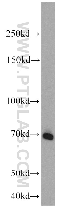
Supportive validation
- Submitted by
- Proteintech Group (provider)
- Main image

- Experimental details
- A549 cells were subjected to SDS PAGE followed by western blot with 10886-1-AP(SDC3 antibody) at dilution of 1:1000
- Sample type
- cell line
Supportive validation
- Submitted by
- Proteintech Group (provider)
- Main image
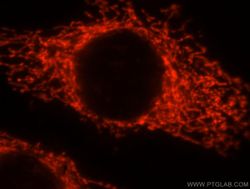
- Experimental details
- Immunofluorescent analysis of MCF-7 cells, using SDC3 antibody 10886-1-AP at 1:25 dilution and Rhodamine-labeled goat anti-rabbit IgG (red).
- Sample type
- cell line

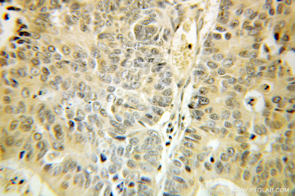
Supportive validation
- Submitted by
- Proteintech Group (provider)
- Main image
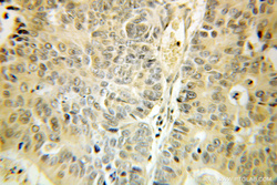
- Experimental details
- Immunohistochemical of paraffin-embedded human colon cancer using 10886-1-AP(SDC3 antibody) at dilution of 1:100 (under 25x lens)
- Sample type
- tissue