 Explore
 Validate
 Learn
Learn Western blot
 Immunocytochemistry
Antibody data
- Antibody Data
- Antigen structure
- References [1]
- Comments [0]
- Validations
- Immunocytochemistry [4]
- Immunohistochemistry [1]
- Other assay [1]
Submit
Validation data
Reference
Comment
Report error
- Product number
- PA5-100116 - Provider product page

- Provider
- Invitrogen Antibodies
- Product name
- Syndecan 3 Polyclonal Antibody
- Antibody type
- Polyclonal
- Antigen
- Synthetic peptide
- Description
- Antibody detects endogenous levels of total Syndecan-3.
- Reactivity
- Human, Mouse, Rat
- Host
- Rabbit
- Isotype
- IgG
- Vial size
- 100 μL
- Concentration
- 1 mg/mL
- Storage
- -20°C
Submitted references Hypoxia Promotes Syndecan-3 Expression in the Tumor Microenvironment.
Prieto-Fernández E, Egia-Mendikute L, Bosch A, García Del Río A, Jimenez-Lasheras B, Antoñana-Vildosola A, Lee SY, Palazon A
Frontiers in immunology 2020;11:586977
Frontiers in immunology 2020;11:586977
No comments: Submit comment
Supportive validation
- Submitted by
- Invitrogen Antibodies (provider)
- Main image
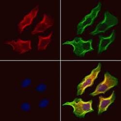
- Experimental details
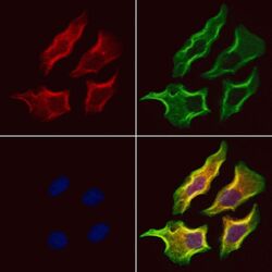
- Immunofluorescent analysis of Syndecan 3 in HeLa cells. Samples were fixed with paraformaldehyde, permeabilized with 0.1% Triton X-100, blocked with 10% serum (45 min at 25°C), incubated with mouse anti-beta tubulin and Syndecan 3 polyclonal antibody (Product # PA5-100116) using a dilution of 1:200 (1 hr, 37°C), and followed by goat anti-rabbit IgG Alexa Fluor 594 (red) and goat anti-mouse IgG Alexa Fluor 488 (green).
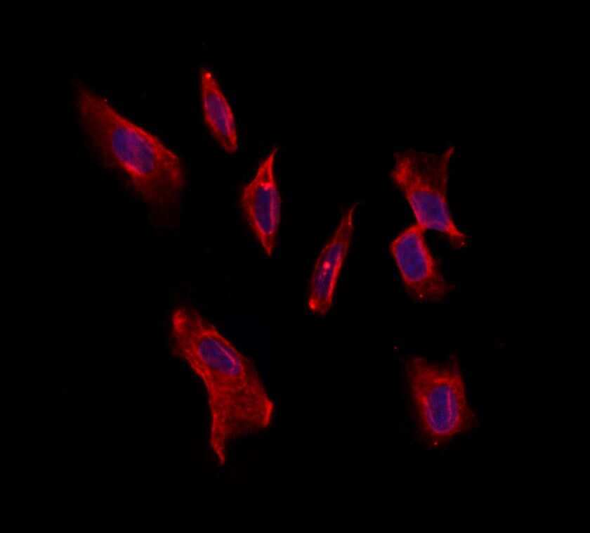
- Submitted by
- Invitrogen Antibodies (provider)
- Main image
- Experimental details
- Immunofluorescent analysis of Syndecan 3 in HeLa cells. Samples were fixed with paraformaldehyde, permeabilized with 0.1% Triton X-100, blocked with 10% serum (45 min at 25°C) incubated with Syndecan 3 polyclonal antibody (Product # PA5-100116) using a dilution of 1:200 (1 hr, 37°C), and followed by goat anti-rabbit IgG Alexa Fluor 594 at a dilution of 1:600.
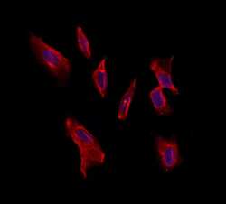
- Submitted by
- Invitrogen Antibodies (provider)
- Main image

- Experimental details
- Immunofluorescent analysis of Syndecan 3 in HeLa cells. Samples were fixed with paraformaldehyde, permeabilized with 0.1% Triton X-100, blocked with 10% serum (45 min at 25°C) incubated with Syndecan 3 polyclonal antibody (Product # PA5-100116) using a dilution of 1:200 (1 hr, 37°C), and followed by goat anti-rabbit IgG Alexa Fluor 594 at a dilution of 1:600.

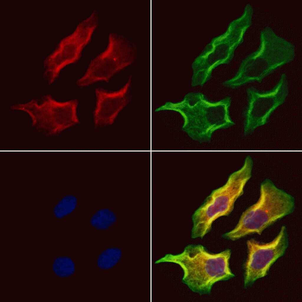
- Submitted by
- Invitrogen Antibodies (provider)
- Main image
- Experimental details
- Immunofluorescent analysis of Syndecan 3 in HeLa cells. Samples were fixed with paraformaldehyde, permeabilized with 0.1% Triton X-100, blocked with 10% serum (45 min at 25°C), incubated with mouse anti-beta tubulin and Syndecan 3 polyclonal antibody (Product # PA5-100116) using a dilution of 1:200 (1 hr, 37°C), and followed by goat anti-rabbit IgG Alexa Fluor 594 (red) and goat anti-mouse IgG Alexa Fluor 488 (green).

Supportive validation
- Submitted by
- Invitrogen Antibodies (provider)
- Main image
- Experimental details
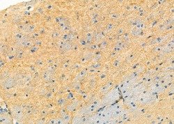
- Immunohistochemistry analysis of Syndecan 3 in mouse brain tissue. The sample was formaldehyde fixed and a heat mediated antigen retrieval step in citrate buffer was performed. Samples were incubated with Syndecan 3 polyclonal antibody (Product # PA5-100116) using a dilution of 1:100 (4°C overnight) followed by HRP conjugated anti-Rabbit secondary antibody.

Supportive validation
- Submitted by
- Invitrogen Antibodies (provider)
- Main image
- Experimental details
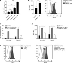
- FIGURE 5 Hypoxia and IFN-gamma drive Sdc-3 expression on tumor associated macrophages. (A) Relative gene expression of Sdc-3 on macrophages derived from murine RAW-264.7 cells treated with increasing doses of DMOG (0, 0.1, 1, and 2 mM) under normoxia (21% oxygen) for 24 h. One representative experiment is shown, error bars represent SD (one-way ANOVA test). (B) Sdc-3 protein expression on macrophages derived from RAW-264.7 cells treated with 1 mM DMOG or vehicle control measured by flow cytometry. One representative experiment is shown, error bars represent SD (unpaired T test), n = 3. A representative histogram showing the geometric Mean Fluorescent Intensity (gMFI) values for each peak is also presented. (C) Relative gene expression of Sdc-3 on macrophages treated with IFN-gamma (100 ng/mL) or IL-4 (100 ng/mL) under normoxia or hypoxia (1% oxygen). One representative experiment is shown, error bars represent SD. Asterisks indicate the p values of the unpaired T test between each condition (normoxia vs. hypoxia). (D) Flow cytometry histograms showing the expression of Sdc-3 protein on macrophages treated with IFN-gamma (100 ng/mL) or IL-4 (100 ng/mL) under normoxia or hypoxia. A pool of two independent experiments is shown, error bars represent SEM (two-way ANOVA test). (E) Representative flow cytometry histograms of Sdc-3 expression on macrophages under normoxia or hypoxia (left) or treated with IFN-gamma (100 ng/mL) or IL-4 (100 ng/mL) under normoxia (right); gMFI values c
